



Retina specialists have been slowly becoming more comfortable treating patients with geographic atrophy (GA) since the US Food and Drug Administration (FDA) approvals of pegcetacoplan (Syfovre, Apellis/Biogen) and avacincaptad pegol (Izervay, Astellas) in 2023. At first, many of us took a cautious approach to integrating these drugs into our clinical algorithms as we waited to see how real-world patients would react to treatment. In 2026, we are treating patients with a wider variety of clinical presentations and, importantly, we are treating them earlier.
To learn more about how and why my colleagues have embraced treatment of early GA, I sat down with Murtaza Adam, MD, and Carl J. Danzig, MD, for a roundtable discussion. We shared real-world cases from our clinics that illustrated the value of early intervention and recorded 3 podcast episodes where we challenged each other on how to treat a series of hypothetical patients whose “real-world” circumstances complicated easy clinical decision-making. Head to Eyetube.net to see our case presentations and listen to New Retina Radio to hear our perspectives. You’ll find ways to directly access the videos and podcasts scattered throughout this discussion.
The roundtable below is adapted from our various discussions and has been edited for brevity and clarity.
— Geeta Lalwani, MD
▶ Want to watch Drs. Lalwani, Danzig, and Adam share real-world clinical cases? Click here.
♫ Listen to Drs. Lalwani, Danzig, and Adam discuss how they would treat specific complicated scenarios on New Retina Radio. Click here.
Imaging Biomarkers, Preferred Imaging Modalities, and Unmet Prognostic Needs
Geeta Lalwani, MD: Use of imaging biomarkers in patients with geographic atrophy (GA) guide our treatment decisions, but it might be difficult to pick a starting point. Which image modality do you first use when seeing a new GA patient?
Carl J. Danzig, MD: Optical coherence tomography (OCT) images are my go-to option. I analyze images for the hypertransmission defect that is a hallmark of GA. This modality is non-invasive, easy to capture, and allows me to use the characterization framework established by the Classification of Atrophy Meeting (CAM) study group, which categorizes retinal atrophy as seen on OCT as incomplete retinal pigment epithelium (RPE) and outer retinal atrophy (also called iRORA) or complete RPE and outer retinal atrophy (also called cRORA).1 This helps me determine how far along GA is in an efficient way.
That said, multimodal imaging approaches serve our patients best, as disease is most thoroughly characterized when we have more than one datapoint. I also send new GA patients for fundus autofluorescence (FAF) imaging, where I can detect lesion patterns (eg, multifocal lesions, diffuse-trickling patterns) associated with rapid disease growth.
A small but important note: I suggest bilateral OCT and FAF imaging for new AMD patients, regardless of the stage of disease. You never know what lurking disease you’ll find in a fellow eye.
Murtaza Adam, MD: FAF imaging helps me assess a patient’s risk of progression. A patient with a unifocal circular lesion 1500 µm from the fovea and no hyperautofluorescence at the lesion border should be treated differently than a patient whose FAF image contains autofluorescent activity at the edge of a large foveal-threatening lesion.
Dr. Lalwani: Does your approach to imaging change after you initiate treatment?
Dr. Danzig: I still order OCT imaging at every visit, as I need to make sure that the patient hasn’t developed concomitant neovascular age-related macular degeneration (AMD). I order FAF imaging, which is less comfortable for patients compared with OCT imaging, approximately every 6 months after they are an established patient. Tracking patient progress on FAF is key to understanding their disease.
Dr. Adam: We are lucky to have these advanced imaging modalities, and we all wish we could use them to help illustrate the effects of intervention with patients in real time. For example, if we could demonstrate that treatment had slowed GA growth toward the foveal center, it would encourage our patients to stick with therapy, and may even sway retina specialists who remain skeptical of complement inhibitors’ efficacy.
Dr. Danzig: Contrast this with imaging in wet AMD, where we can show patients how treatment has resulted in fluid resolution and restoration of anatomy.
A technology that shows patients the effects of intervention in GA seems like something that would be conducive to an AI-powered application. If I could show patients an image of how their disease would most likely have progressed without treatment and compare it to the image captured in clinic, I think they would understand the value of treatment. An AI forecast of how a patient’s disease would progress could tell us a lot about the need to intervene, too.
This could also allow us to catch patients further upstream of GA. If a patient with intermediate AMD presents to the clinic, perhaps AI-powered imaging could tell us how quickly and in what way they’re likely to progress to advanced AMD.
“Starting treatment now is like investing money when you’re young—the earlier you invest, the more robust your returns.”
-Dr. Adam
Increased Comfort with Early Intervention
Dr. Lalwani: It is my impression that retina specialists have become more comfortable with treating GA earlier in the disease process. My personal clinical practice reflects this. Have you noticed trends in the field or in your clinic that differ from a year or 2 ago?
Dr. Adam: Your observations match mine. When complement inhibitors first made an appearance in my clinic, I was cautious: as reliable as clinical trial data are, I still wanted to see how real-world patients would react to treatment. As my experience with complement inhibitors has increased, so too has my confidence that I have selected the correct patients for an optimal outcome. This means that I’m treating more patients and treating them earlier. I am now taking a much more proactive approach with younger patients with early GA, whereas a few years ago, I may have taken a wait-and-see approach to treating such patients.
Dr. Lalwani: For me, knowing that GA can progress to foveal involvement within 2.5 years challenges our previous characterization of GA as a slow-moving disease.2 These data confirm something many of us already saw on FAF imaging in our clinics.
Dr. Danzig, how have your practice patterns changed?
Dr. Danzig: I enthusiastically embraced complement inhibition when we first had access to it in 2023—but I also proceeded with caution, like Dr. Adam. Patients with advanced disease were the first to receive this treatment in my clinic. However, the more I saw GA patients, the more comfortable I became with injecting earlier in the disease course.
Today, I regularly treat patients with good vision and extrafoveal lesions. Even in patients where I still think it’s too early to treat—say, in a patient with a very small extrafoveal, unifocal lesion—I schedule them for a 4- to 6-month follow-up appointment rather than a 9- to 12-month follow-up appointment.
Dr. Adam: There is a temptation to treat patients only if they would have qualified for enrollment in one of the phase 3 clinical trials that assessed the safety and efficacy of avacincaptad pegol (Izervay, Astellas) or pegcetacoplan (Syfovre, Biogen). But that overlooks that clinical trials are designed to be efficient and to reach a regulatory endpoint, which means that they did not necessarily include many of the patient types we see in practice.
The fact remains that patients who would not have qualified for a phase 3 clinical trial nevertheless have common risk factors linked with faster progression. When a patient has such risk factors, I educate them about the specifics of their GA so that they can make an informed decision. For example, a patient with multifocal lesions more than 1500 µm from the foveal center is, in my estimation, going to benefit from treatment, even if the distance of their disease from the foveal center falls beyond the typical enrollment criteria of a clinical trial.
Dr. Danzig: Our practice patterns have certainly changed, which is in part due to the change in referral patterns. Optometrists and referring ophthalmologists are better able to detect GA and intermediate AMD, and they have started to refer patients to our clinic earlier in the disease course now that they are educated about the value of treatment.
Retina clinics offering photobiomodulation therapy (Valeda Light Delivery System, Alcon) may attract referrals from providers interested in intermediate AMD treatment, which means that those intermediate AMD patients will already be under our care. When we detect nascent GA in those patients, we’ll be able to treat them earlier. Getting patients integrated into the clinic via photobiomodulation may ultimately be an unlock for early GA treatment, as many of them would not have presented or been referred to our clinics until they were symptomatic—which may, in some cases, mean that their foveal anatomy was already compromised.
Dr. Adam: I emphasize early referrals with my referral base. When complement inhibitors were first approved, many referring providers referred patients with significant GA and VA 20/200 or worse. We could not do much for these patients. After diligent educational efforts, our referring providers send patients with earlier disease for consultation, increasing the likelihood that we can help them.
Further, when we help patients with early disease, we may be buying them time until a more effective therapy is approved. If we can slow the growth of GA lesions for several years ahead of a novel treatment that either slows it more, arrests it, or even restores some vision, then we’ve done our patients a major service.
Dr. Lalwani: Which other factors play into the decision to treat GA?
Dr. Danzig: Treatment relies heavily on a constellation of datapoints, not all of which are captured on imaging. The patient’s overall health profile and their motivation to commit to treatment are among the biggest factors I consider.
When I discuss treatment with a patient who says they’re open to it, I always begin by saying that the label articulates monthly dosing for avacincaptad pegol and monthly or every-other-month dosing for pegcetacoplan. I prefer to start with monthly treatment, and if they agree that regimen is realistic and sustainable, then I feel confident that, if they elect treatment, that they will be a good candidate for long-term therapy.
Dr. Adam: Patients sometimes come to the clinic highly educated and ready to select a treatment. In those cases, take yes for an answer.
I try to keep it simple. If they want avacincaptad pegol, then I begin treatment with avacincaptad pegol and avoid talking them into pegcetacoplan, or vice versa. Both drugs are safe and effective, and I am confident that we are equipped to manage any potential adverse events. In short, if an educated patient wants treatment, then treat.
Of course, if there is some reason to choose one drug over the other given that patient’s specific circumstances, then use your clinical judgement.
Dr. Lalwani: Describe the benefits of beginning with monthly therapy.
Dr. Danzig: If a patient starts on monthly treatment and finds that it’s too difficult to maintain, we can adjust to treating every 6 weeks, which still leaves a 2-week buffer to every-other-month treatment. That buffer comes in handy when life events or clinical schedules mean a patient will be a week or two late.
However, if you start with every-other-month treatment, the patient is very unlikely to request more frequent treatment. If they miss an appointment or are otherwise unable to attend clinic, they’re now receiving less than the recommended dosage of therapy, which means they may experience diminishing returns.
We should note that avacincaptad pegol is approved for monthly dosing, and pegcetacoplan is approved for monthly or every-other-month treatment.
Dr. Lalwani: Conversations around treatment regimen are one thing; conversations about disease education are another. How do you educate the patients with early GA—who may not experience any subjective disruption to their vision—that their condition is worthy of treatment?
Dr. Adam: I start with a simple fact: that they are at risk of losing vision within the next several years. Even if they don’t have risk factors associated with rapid progression, they still have a GA lesion, and natural history studies suggest that this lesion will grow.
I don’t push for same-day treatment. I understand that the volume and weight of the information I just gave a first-time GA patient is a lot to process. I take the time to show them examples of GA and show them 6-month results with and without treatment. I also make sure to show my patients with GA the evolution of their own imaging over 4 to 6 months, if they choose not to start treatment soon after our first consultation. We can detect differences even in that short window, and images of growth are easy to understand.
Dr. Danzig: Many patients come with a caregiver, such as a spouse, family member, or friend. They can serve as a second set of ears for many patients, which is especially useful at the time of a diagnosis. They can also be someone who encourages the patient to undergo treatment, as they’ll explain that helping drive them to appointments is an obligation they’re happy to take on.
One logistical pearl that has helped my clinical workflow: ask patients before you begin explaining GA to them if they have someone in the waiting room who could benefit from participating in this conversation. If they do, bring that person into the examination room so they can both be present for the educational session.
I’ve experienced appointments where I spend significant chair time explaining the disease and treatment options, only for the patient to ask if they can now bring their caregiver into the room so I can explain it again. I am happy to do that, but I find that if they’re both present for the conversation, I can recoup valuable time.
Dr. Lalwani: Extension studies for both drugs have shown that there is a compounding effect of the drug over time, meaning that the longer a patient is undergoing treatment, the wider the differential between a treated eye and an untreated eye.3,4 How do you explain this concept to patients?
Dr. Adam: I speak in terms of saving money, where the concept of compounding interest is relevant. I explain how starting treatment now is like investing money when you’re young—the earlier you invest, the more robust your returns.
Dr. Lalwani: Our field was previously uninterested in treating younger patients and patients with earlier disease, but as we’ve noted, that dynamic is changing. How do you approach treating younger patients?
Dr. Danzig: I practice in Florida, where the average age is higher than the rest of the country, so my definition of young may include a patient in their 80s with an active lifestyle and otherwise good health. The age of the patient is less relevant in GA than the duration of disease, and I think retina specialists have begun to value early treatment in younger patients given the compounding phenomenon described above.

Real-World Case: Delayed Complement Inhibition Therapy Per Patient Request
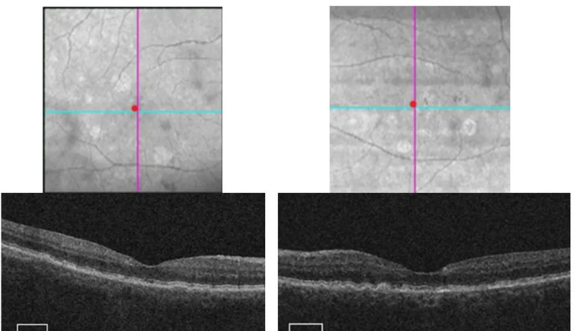
Figure 1. SD-OCT imaging shows bilateral extrafoveal GA lesions, but no hypertransmission defects. VA is 20/20+2 OD and 20/25+1 OS.
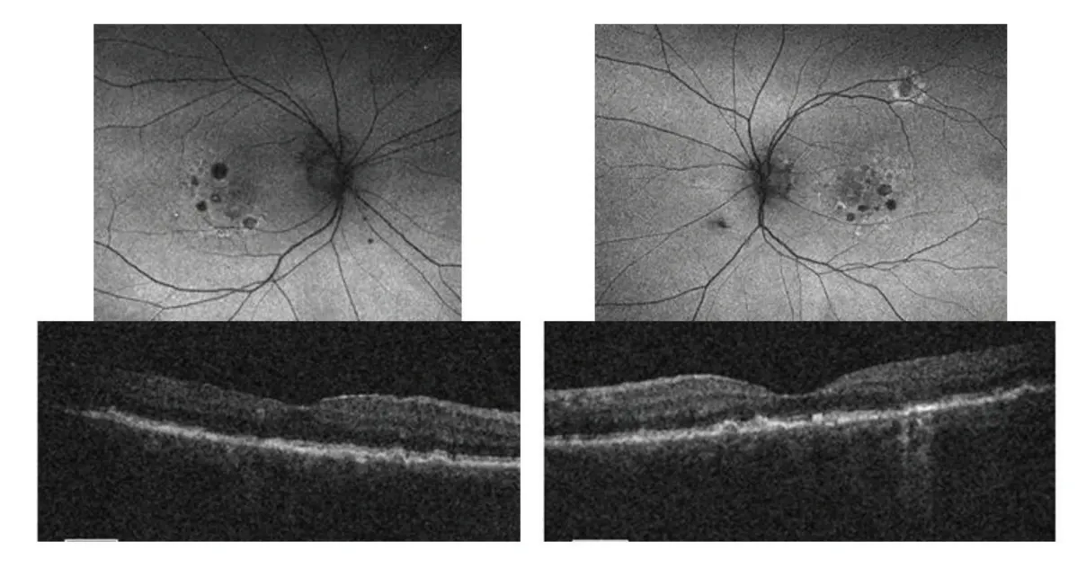
Figure 2. Small extrafoveal lesions are observed bilaterally on FAF imaging, and hypertransmission is observed on OCT in the left eye. Vision declined to 20/25-2 OD and 20/20-2 OS.
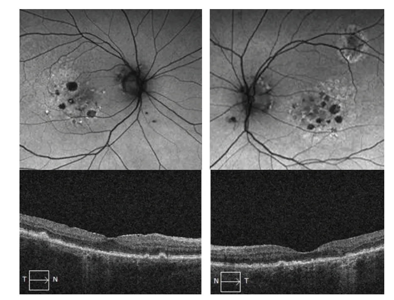
Figure 3. Eleven months later, the patient returned with decreased visual acuity, measured at 20/30-2 OD and 20/30 OS. Bilateral extrafoveal lesion growth was observed on FAF and hypertransmission remained visible on OCT. The patient opted to begin complement inhibition.

Dr. Lalwani: How do you approach conversations around therapy for a patient who is a good candidate for complement inhibition but is reluctant to start treatment?
Dr. Adam: Patients are often overwhelmed when they hear their diagnosis, and many of them are distracted by the fear of going blind. And we must remember that 40% to 80% of information presented during an office visit is misremembered or forgotten by the patient, and that patients forget more as we present a higher volume of information.5
In a case such as this one, I would probably stick to educating the patient about their condition and their treatment options, trusting that factual repetition will result in a properly educated patient. Your use of images to educate this patient was wise: we can talk all we want to about concepts surrounding GA, but as soon as we show patients pictures, they grasp the concepts they need. After they understand the basics of GA, I often tell them that they have two options: to sit on their hands and wait, or to intervene early in hopes of retarding growth.
We shouldn’t overlook the very useful print literature that Astellas and Biogen provide. These resources can be reviewed by patients and caregivers at their own pace, act as references for when patients may have questions at home, and serve as useful reminders that they should follow up with their provider.
Dr. Danzig: If a patient with bilateral disease is open to (but nevertheless worried about) treatment, I emphasize to them that we’ll begin with one eye so that they can get used to treatment. I would probably only start with unilateral injections anyway when beginning complement therapy to assess the patient’s response to treatment, so this approach accomplishes two objectives—safety testing and slowly introducing the patient to therapy—with a single approach.
For an active patient such as this one, I remind them that treatment can hopefully preserve their independence. The loss of driving privileges could significantly decrease a patient’s quality of life, and the sooner we begin treatment, the longer a patient may be able to safely drive.
Dr. Lalwani: When the patient first presented, I relied only on SD-OCT, as it was the only modality available in my clinic that day. When the patient returned, I included FAF imaging alongside OCT imaging. I found multimodal imaging valuable. But is it always needed, particularly in early cases?
Dr. Danzig: It’s so easy to miss GA in a rapidly moving clinic, and even easier to miss if we only rely on a single modality. When patients first present, we want to capture as much data as possible. We might, for example, see GA lesions on FAF but not see hypertransmission on OCT; in this case, a multimodal approach could be the difference between detection and overlooking disease. Personally, I find GA poorly depicted on color fundus photography and stick to OCT and FAF when possible.
After a patient begins treatment, I stick to OCT imaging at every visit and use FAF imaging only once every 6 months.
Real-World Case: Complement Inhibition Therapy in a Monocular Patient With a New CNV
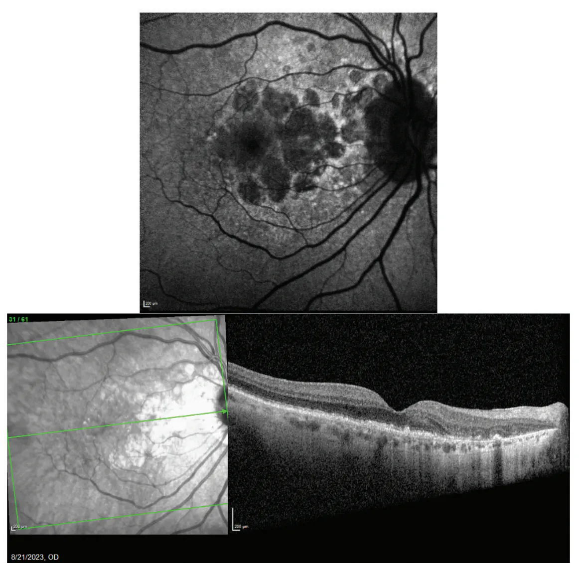
Figure 4. FAF imaging OD revealed large, multifocal, extrafoveal GA lesions. OCT imaging showed hypertransmission close to the fovea (bottom).
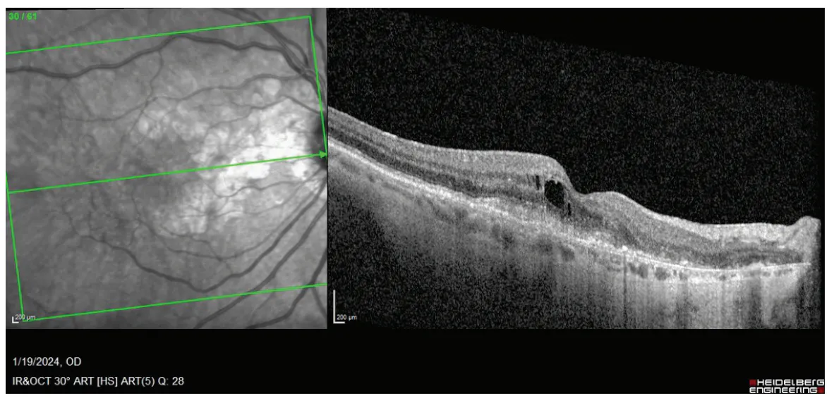
Figure 5. OCT imaging depicts a subfoveal neovascular membrane and foveal edema, which was detected on a follow-up appointment after the patient’s second complement inhibition injection.
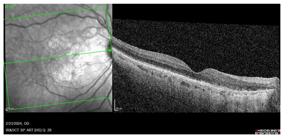
Figure 6. One month after receiving bevacizumab to address her subfoveal neovascular membrane, the patient’s anatomy resolved, even as VA remained 20/40 OD.
Dr. Lalwani: This case illustrates how limited access to care can lead to catastrophic conditions—in this case, a fibrotic scar that left the patient CF OS. How did you explain that her GA OD was different from her wet AMD OS?
Dr. Danzig: I spent less time explaining the differences in pathology than I did explaining how a lack of treatment can lead to permanent visual impairment—a concept the patient subjectively understood given her VA of CF OS. The GA lesions OD were large and various on FAF. Showing the patient this image helped her grasp the severity and advanced nature of her disease.
“Treatment relies on a constellation of datapoints.”
-Dr. Danzig
Dr. Lalwani: What approaches to treating wet AMD concomitant with GA are most often used?
Dr. Adam: First, I commend Dr. Danzig for doing his best to make sure this patient did not fall through the cracks. Given that she is monocular and drives several hours to clinic, it’s easy to see how she could be knocked off course. Getting this patient into a routine with alternating monthly treatment with either anti-VEGF or complement inhibition is key to success.
For a patient without those circumstances, I would try to use a treat-and-extend (TAE) approach for the neovascular disease. Like Dr. Danzig, I pause complement inhibition until neovascular disease is controlled. After sufficient extension of anti-VEGF therapy is reached, we then have the bandwidth to continue GA treatment.
In my experience, neovascular disease occurring in a patient on complement inhibition therapy is often caught early and is mild in nature. Promptly addressing it with anti-VEGF is sufficient for rapid resolution. In some cases, as-needed treatment is more appropriate than a TAE approach, especially if there is no evidence of neovascular disease recurrence over several appointments.
Dr. Danzig: If we lived in a world where insurance issues didn’t dictate treatment decisions, then I would have opted for a next-generation treatment that might allow for longer duration of treatment for her neovascular disease. However, in not wanting to delay care and not wanting to push the patient through a series of insurance hurdles, I chose bevacizumab treatment. Clearly it was sufficient in this case to address neovascular disease.
1. Guymer RH, Rosenfeld PJ, Curcio CA, et al. Incomplete retinal pigment epithelial and outer retinal atrophy in age-related macular degeneration: Classification of Atrophy Meeting report 4. Ophthalmology. 2020;127(3):394-409.
2. Lindblad AS, Lloyd PC, Clemons TE, et al; Age-Related Eye Disease Study Research Group. Change in area of geographic atrophy in the Age-Related Eye Disease Study: AREDS report number 26. Arch Ophthalmol. 2009;127(9):1168-1174.
3. Wykoff CC, Holz FG, Chiang A, et al; OAKS, DERBY, and GALE Investigators. Pegcetacoplan treatment for geographic atrophy in age-related macular degeneration over 36 months: data from OAKS, DERBY, and GALE. Am J Ophthalmol. 2025;276:350-364.
4. Khanani AM. Avacincaptad pegol for GA: 3-year results from the GATHER2 open-label extension trial. Presented at: American Academy of Ophthalmology Annual Meeting; October 17-20, 2025; Orlando, FL.
5. Kessels RP. Patients’ memory for medical information. J R Soc Med. 2003;96(5):219-222.
