


The ZEISS Retina Workflow in the Clinic | By Katherine Talcott, MD
For me, OCT is the diagnostic backbone behind nearly every clinical decision. In particular, multimodal imaging that is available on integrated platforms like the ZEISS Retina Workplace has reshaped how we practitioners identify, monitor, and explain disease to patients. The ability to combine high-resolution structural OCT, fundus autofluorescence, infrared imaging, and composite B-scans within a single workflow has made it possible to assess and treat pathology with much greater clarity and efficiency.
Following Geographic Atrophy
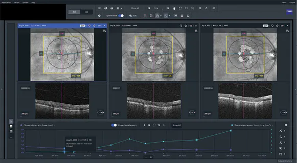
Figure 1. A patient with GA in the right eye as seen with infrared and OCT images showing progression over a 3-year period.
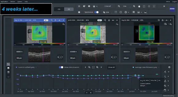
Figure 2. Patient with nAMD on anti-VEGF injections who had worsening subretinal fluid and hemorrhage at 8 weeks (middle panel) that improved 4 weeks later (right panel) after another injection.
A particular case of geographic atrophy (GA) illustrates how I use these tools in my practice. I’ve been following a 76-year-old woman who has had stable 20/25 UCVA OD for the past 3 years. Her fellow eye has count-fingers vision from a longstanding choroidal hemangioma. At her most recent visit, she reported increasing difficulty with reading, one of her favorite activities.
Structural OCT of her right eye showed no exudation, but multimodal imaging revealed the broader picture. On fundus autofluorescence, I saw areas of retinal pigment epithelium (RPE) loss surrounding the fovea, and with infrared imaging I noted the atrophic areas. Composite B-scans gave me additional confirmation that the atrophy in the right eye had expanded toward the fovea. In the left eye, unfortunately, the diffuse atrophy already involved the fovea.
It is very helpful to be able to compare earlier scans from all these modalities on the ZEISS Retina Workplace software. Scans of this patient’s right eye from 2 years prior showed almost no atrophy, but scans from her last 3 visits revealed its progression (Figure 1). The composite B scan images, in particular, I find ideal not only for identifying GA and loss of RPE, but for educating patients so we can discuss the appropriate treatment options.
Anti-VEGF Evaluations
The ZEISS Retina Workplace is also great for evaluating patients who receive anti-VEGF injections. One of my patients with neovascular age-related macular degeneration had undergone 37 injections of aflibercept (Eyelea, Regeneron) in his left eye. We had not been able to extend the injections beyond 5 weeks before the fluid returned. At one point, he was interested in trying a newer anti-VEGF agent, which allowed us to extend to a 6-week interval. The following scan showed a reduction in fluid, and his vision was fine. When we tried an 8-week interval, however, the subretinal fluid returned, even more so than when he was on aflibercept, and I also found a hemorrhage (Figure 2). Ultimately, he decided to switch back to aflibercept at a 6-week interval. This case underscores the degree to which I rely on multimodal imaging to follow these types of patients and decide on the most effective treatment intervals.
OCT-A
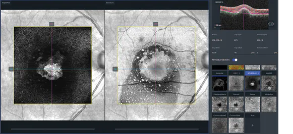
I find myself relying on OCT-angiography (OCT-A) over fluorescein angiography for some diagnoses, especially choroidal neovascular membranes. For example, a 45-year-old patient with chronic central serous chorioretinopathy experienced a sudden decline in vision to 20/60 after years of stability. Structural OCT showed subretinal fluid and subretinal hyperreflective material, which made me suspect secondary choroidal neovascularization. Yet, on OCT-A, I used segmentation to find flow signals beneath the RPE that revealed the presence of a neovascular membrane (Figure 3). We initiated anti-VEGF therapy, which resolved the fluid and preserved the patient’s vision.
ZEISS FORUM
FORUM is ZEISS’ data management software that enriches my understanding of a patient’s imaging and how to manage their retinal disease. Its advanced review tools allow me to quickly synthesize changes in their imaging, including in response to treatment. For example, it tracks and applies time points to central subfield imaging, so that I can quickly compare scans from different dates and identify blips when more fluid was present. This is invaluable when a patient asks to extend the intervals between their injections. I can show them on their imaging history exactly the intervals at which the fluid recurred, and also how far they’ve progressed since starting treatment.
I appreciate having a platform that unifies imaging and diagnostic tools and that is accessible across the clinic and OR.

Katherine E. Talcott, MD
- Retina specialist at the Cleveland Clinic
- talcotk@ccf.org
- Financial disclosure: The authors / speakers have a contractual or other financial relationship with Carl Zeiss Meditec AG and its affiliates and have received financial support.
The ZEISS Retina Workflow in the OR | By Jorge Fortun, MD
In the OR, the ZEISS Retina Workflow is my workhorse. This integrated system includes the EVA NEXUS vitreoretinal and cataract surgical system by DORC (a ZEISS company) and the ARTEVO 850 3D digital microscope by ZEISS. Together, they enable me to perform more advanced surgery, reflective of the shift in our field away from purely traditional vacuum-based vitrectomy consoles and toward a true hybrid model of surgery with improvements in fluidics control, pressure management, visualization, and, ultimately, a more deliberate way to operate.
Ultra-Widefield Imaging and Intraoperative OCT
I have operated with heads-up displays for years, and ZEISS ARTEVO 850 has become my go-to system because it gives me ultra-widefield imaging and intraoperative OCT combined. Having a shared surgical view for everyone in the room, especially when I’m teaching, has been a real advantage. Instead of trying to describe subtle tissue planes through a teaching scope or a secondary monitor, I can walk people through the case as it is happening.
Intraoperative OCT allows me to be that much more responsive during surgery. It gives me real-time diagnostic feedback at the moment when I’m deciding whether to stop, whether to keep peeling, or whether the tissue is behaving the way I think it is. In macular surgery, it helps confirm whether a peel is complete or whether something unexpected has happened. In posterior proliferative vitreoretinopathy, it can show residual tissue that may not be obvious through the microscope alone. In an optic pit case, intraoperative OCT can show in real time how much improvement we have achieved after vitrectomy and hyaloid elevation, which may spare the patient unnecessary additional maneuvers like internal limiting membrane (ILM) peeling.
Also, I think that intraoperative OCT is going to become increasingly important as we move further into subretinal therapies and revisit submacular surgery. The more our maneuvers depend on exact tissue-plane information, the more valuable it is to have OCT integrated directly into the surgical workflow rather than relying on inference alone.
One case that captures this well was a surgery for an optic pit my partner Luis Haddock, MD, performed. These eyes always raise the question of how much intervention is needed, because every additional step carries risk. In that case, intraoperative OCT showed meaningful improvement after vitrectomy and hyaloid elevation alone. That real-time feedback helped guide the case and made it clear that more intervention, including ILM peeling, was not necessarily required. To me, that is exactly where this technology proves its value: in helping us make better intraoperative decisions.
ZEISS Resight
The ZEISS RESIGHT 500/700 fundus viewing system is another place where the practical value shows up every day. The Single-use noncontact Lenses for ZEISS RESIGHT, including the ULTRA WIDE-ANGLE Lens, provide practical flexibility and excellent optics. The wider peripheral view can reduce the need for scleral depression in selected cases, which is particularly helpful early in practice or in settings without highly experienced assistance. For surgeons building an ambulatory surgery center, disposability also offers real-world advantages in cost structure, turnover, and optical consistency.
Conclusion
The ZEISS Retina Workflow offers a true value proposition: ZEISS ARTEVO 850 for a better heads-up surgical view, intraoperative OCT for real-time decision-making, and the ZEISS RESIGHT fundus viewing system for efficient, flexible visualization from the macula to the far periphery. These tools fit naturally into the way we already operate, but they let us do it with more information and, I would argue, with more confidence. n

Jorge Fortun, MD
- Vitreoretinal surgeon at Bascom Palmer Eye Institute, Miami
- jfortun@med.miami.edu
- Financial disclosure: The authors / speakers have a contractual or other financial relationship with Carl Zeiss Meditec AG and its affiliates and have received financial support.
en-INT_31_200_0414I The statements of the authors / speakers reflect only their personal opinions and experiences and do not necessarily reflect the opinion of any institution with whom they are affiliated that they are affiliated with. The authors / speakers alone are responsible for the content of their experience reported and any potential resulting infringements. Carl Zeiss Meditec AG and its affiliates do not have clinical evidence supporting the opinions and statements of the authors / speakers nor accept any responsibility or liability of the authors’/speakers' content.
DORC EVA NEXUS are either trademarks or registered trademarks of Carl Zeiss Meditec AG or other companies of the ZEISS Group in Germany and / or other countries. Products referenced may not be available in all countries and product labeling varies by country.
