


Case Presentation
A 28-year-old man was referred to us for repair of a traumatic macular hole with subretinal fibrosis. He had central blurry vision after sustaining a punch to the left eye six years ago. His presenting visual acuity was counting fingers at 1 foot. The surgical plan was to perform a vitrectomy with peeling of the internal limiting membrane (ILM), using the ILM flaps to cover the hole, followed by placement of an amniotic membrane graft to augment hole closure and silicone oil tamponade.
Amniotic Membrane Placement Facilitated By Intraoperative OCT
Because we were using an amniotic membrane in this case, we used 23-gauge instrumentation with a twin light chandelier so we could operate with a bimanual approach. Furthermore, we performed the surgery using the EVA NEXUS surgical platform (DORC, a ZEISS company) and the ARTEVO 850 visualization system (ZEISS), each of which played an important role in the success of the surgery.
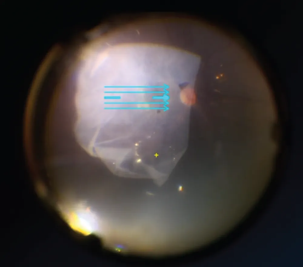
For the surgery, we started with vacuum mode, which allowed for a fast and efficient core vitrectomy; flow mode was used for precise aspiration closer to the macular hole. A blue filter on the ZEISS ARTEVO 850 was used to aid visualization of the injected intraoperative dye. Next, we placed a large amniotic membrane graft that covered the posterior pole followed by silicone oil.
CPT Code: 67042 (Vitrectomy, pars plana approach; with removal of internal limiting membrane of retina)
Intraoperative OCT was used for several parts of the procedure. During ILM peeling, it helped confirm that the ILM was peeled to the edges of the hole and the flaps were in the proper position overlying the hole. At the end of the surgery, intraoperative OCT was used to ensure proper placement of the amniotic membrane.
At postoperative month 3, the silicone oil was removed. The patient’s vision improved to 20/400. At the most recent follow-up, the macular hole remained closed.
Discussion
Complex macular hole repair requires the use of advanced vitreoretinal surgical techniques. Furthermore, although the steps of the procedure may be planned in advance, there is often a need to adjust intraoperatively based on the anatomy. These factors highlight the importance of using a surgical platform that enhances the surgeon’s ability to perform safe and efficient maneuvers in the back of the eye, and a visualization system that allows a clear view of the retina and its structures.
Fundamentally, intraoperative OCT has two advantages: it helps avoid unnecessary movements and cutting, and it greatly enhances decision-making to do what is necessary. In the not-so-distant past, intraoperative OCT was considered a “nice to have” feature during vitreoretinal surgery. However, as the field continues to advance, and as the demand on the surgeon to be precise while maneuvering delicate tissue enhances, the utility of intraoperative OCT is becoming more apparent. In this case, real-time viewing allowed us to confirm that the ILM had been peeled to our satisfaction, and that no remnant pieces were left behind. At the end of the case, we were able to confirm the placement of the amniotic membrane before leaving the OR.
Digital visualization with the ARTEVO 850 has a couple of features that were pertinent to this case. For instance, we were able to switch seamlessly between 3D viewing on the monitor and ocular viewing with the microscope—a feature that is not commonly available on other heads-up display systems. As well, the use of light filters facilitated precise views of the various retinal tissues; in this case, intraoperative dye highlighted areas of ILM requiring peeling, and the addition of a blue filter further enhanced the view. Another consideration is that because we performed most of the case in digital view, we were using reduced luminance, thereby reducing the potential for phototoxicity.
