




Please see link to Prescribing Information here.
An Introduction by Apellis
More than 3 years have passed since SYFOVRE® (pegcetacoplan injection) 15 mg/0.1mL received approval from the US Food and Drug Administration (FDA) for the treatment of geographic atrophy (GA) secondary to age-related macular degeneration (AMD), marking the first therapy approved for GA. In the Phase 3 OAKS and DERBY trials, pegcetacoplan demonstrated a reduction in the rate of GA lesion growth with over 24,000 injections in the clinical trial setting.1 As of May 2026, over 1 million injections of pegcetacoplan have been administered.2*
Pegcetacoplan efficacy and safety were assessed in OAKS (N=637) and DERBY (N=621), multicenter, 2-year, Phase 3, randomized, double-masked trials. Patients with GA secondary to AMD with or without subfoveal involvement were randomly assigned (2:2:1:1) to receive 15 mg/0.1 mL intravitreal pegcetacoplan monthly, or every other month (EOM), sham monthly, or sham every other month for 2 years. Change from baseline in rate of GA lesion area growth in the study eye (mm2) was measured by FAF.1 Safety data from OAKS and DERBY is summarized in Table 1.
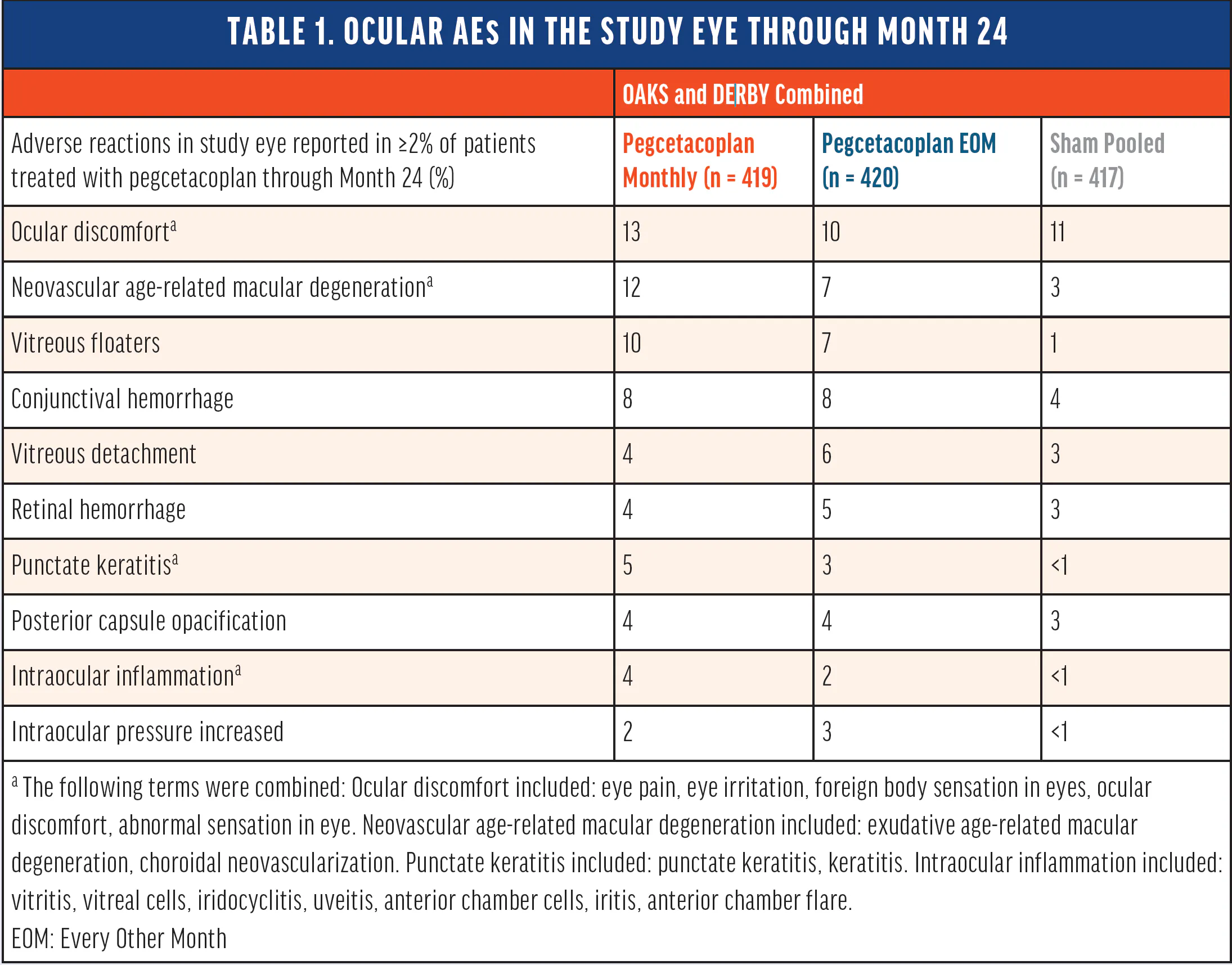
At Year 2, pegcetacoplan reduced GA lesion growth by 18% and 22% with monthly dosing and 17% and 18% with EOM dosing vs sham pooled.1
Following FDA approval, cases of retinal vasculitis were reported with real-world use. Apellis, a subsidiary of Biogen, responded promptly, working with the American Society of Retina Specialists (ASRS) Research and Safety in Therapeutics (ReST) Committee in an effort to ensure transparent communication with the retina community. Drug safety and pharmacovigilance remain central to Apellis’ values, prompting a comprehensive evaluation of these events in an effort to better understand their causes and outcomes.
It is important to note retinal vasculitis and/or retinal vascular occlusion, typically in the presence of intraocular inflammation, have been reported with the use of SYFOVRE. Cases may occur with the first dose of SYFOVRE and may result in severe vision loss. Discontinue treatment with SYFOVRE in patients who develop these events. Patients should be instructed to report any change in vision without delay.1
Could you provide an overview of Apellis’ process for adjudication and evaluation of potential retinal vasculitis cases?
- Retinal vessel leakage observed on FA
- Optic nerve leakage observed on FA
- Retinal vascular occlusion observed on FA (for occlusive cases only)
- Vessel sheathing on fundus photography
Apellis submits all reported adverse events to the FDA and other applicable Regulatory Agencies consistent with reporting guidelines and regulations for drug manufacturers.
How many confirmed cases of vasculitis have there been since 2023?
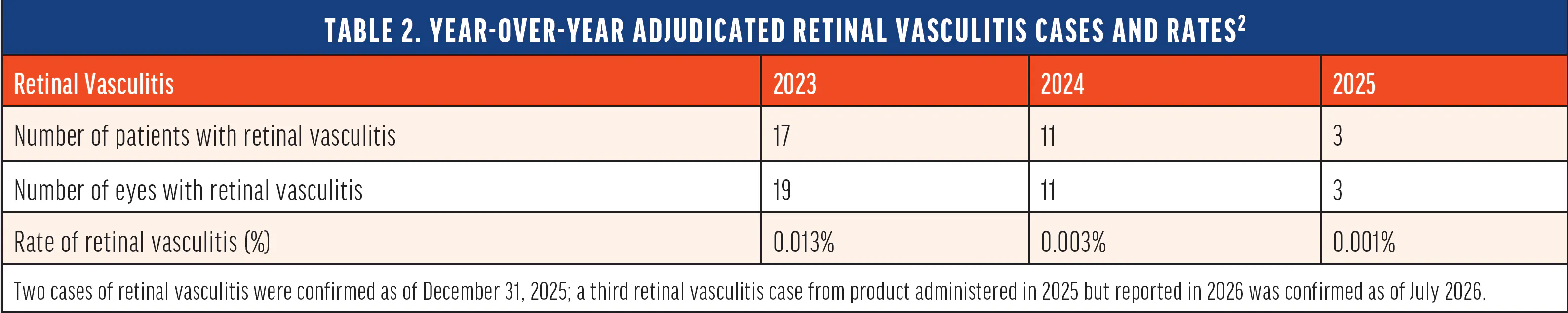
Since approval, the annual reporting rate of eyes adjudicated as having been diagnosed with vasculitis subsequent to the use of SYFOVRE was 0.013% in 2023, 0.003% in 2024, and 0.001% in 2025 (Table 2). Please note that the post-marketing data on retinal vasculitis is current as of December 2025.
As of December 2025, more than 133,000 patients had received pegcetacoplan injections. The incidence rate of vasculitis per first injection is approximately 1 in 4,000 (0.025%). The estimated rate of retinal vasculitis with severe vision loss is approximately 1/11,000 per first injection (0.0091%). Severe vision loss is defined as ≥6 lines lost (Table 3).2
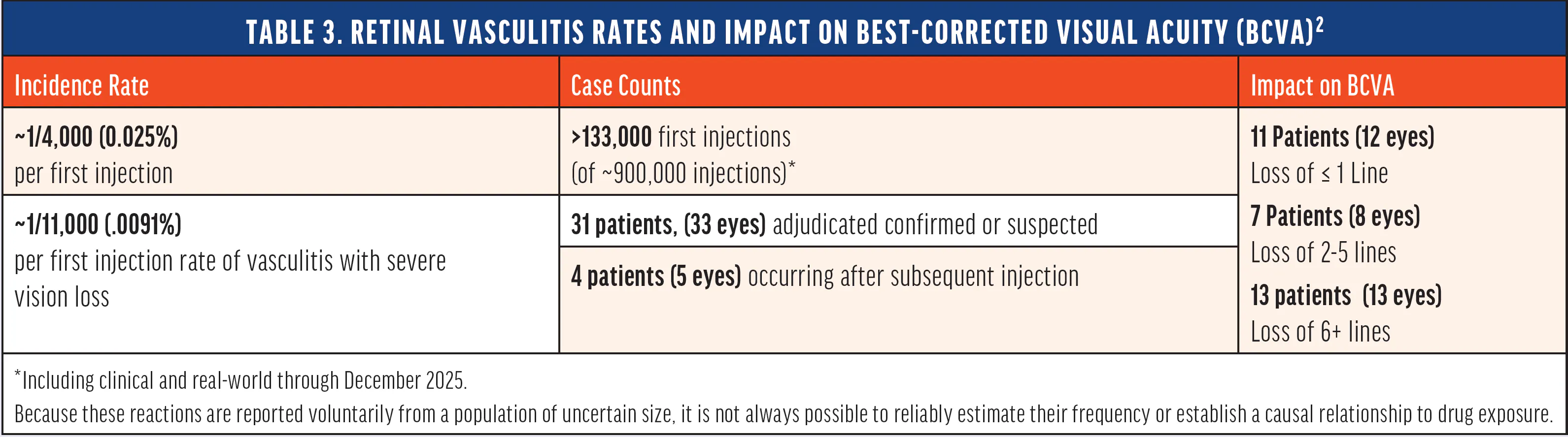
Retinal vasculitis has been adjudicated in 31 patients (33 eyes).2
- 11 patients (12 eyes) had a visual acuity recovery to within ≤1 line of baseline
- 7 patients (8 eyes) had a loss of 2-5 lines
- 13 patients (13 eyes) had a loss of ≥6 lines at last reported follow up
Although post-marketing data suggests retinal vasculitis is primarily a first-injection phenomenon, 4 patients (5 eyes) developed vasculitis following subsequent injections (Table 3). Of these eyes, 4 eyes returned to baseline vision after treatment. As of December 2025, more than 760,000 subsequent injections have been administered. The overall estimated rate of retinal vasculitis with subsequent injections is approximately 1 in 190,000 (~0.0005%).2
Real-world data from the IRIS® Registry indicate that retinal vasculitis occurs at a rate of approximately 1-2 cases per 100,000 anti-VEGF intravitreal injections in patients with wet AMD and GA.4
If vasculitis is suspected after pegcetacoplan use, report the case to Apellis Safety (via Med Info):
- Phone: 1-833-866-3346
- Email: medinfo@apellis.com
- Minimum information to be included:
- Patient identifier (e.g., date of birth, age, gender)
- Description of event(s)
- Apellis product involved
- Reporter contact information
1. Syfovre. Package insert. Apellis Pharmaceuticals, Inc.; 2025
2. Data on file. Apellis, A Subsidiary of Biogen
3. Albini TA, Yeh S, Regillo CD, et al. Expert opinion: management of intraocular inflammation and vasculitis after intravitreal pegcetacoplan for geographic atrophy. Ophthalmic Surg Lasers Imaging Retina. 2026:1-7.
4. Leng T, Schneider EW, Rahimy E, et al. IRIS® Registry analysis of anti-VEGF treatment in patients with coexisting neovascular age-related macular degeneration and geographic atrophy. Oral presentation at: The Association for Research in Vision and Ophthalmology Annual Meeting; May 5-9, 2024; Seattle, Washington.
3 Years After Pegcetacoplan Approval: Addressing Post-Injection Complications and Understanding EZ:RPE Loss as a Potential Measure of Clinical Response
A panel of leading experts shares their experiences managing one of the most time-sensitive complications of pegcetacoplan injection before turning to emerging data from AI-based imaging analyses that are enhancing our understanding of GA disease progression.
There is no standard protocol for treating retinal vasculitis following an intravitreal injection. In the interest of hearing how leading retina specialists detect, treat, and follow patients who present with these post-injection complications, Apellis convened a roundtable where retina specialists could share their ideas and approaches. This article presents the highlights of that discussion.
The roundtable begins with a case published by Leung et al that describes a real-world patient whose vision was restored following an occlusive retinal vasculitis event occurring after treatment with pegcetacoplan injection that temporarily reduced her vision to counting fingers.1 The panel discusses the particulars of that case and uses it as a backdrop to discuss other issues at play in modern practice before moving to a wider discussion about the utility of next-generation biomarker-based risk stratification.
The discussion around treating retinal vasculitis may be particularly useful for some readers. The retina community has expanded its retinal vasculitis knowledge base, and this roundtable presents the opportunity to see how world-class retina specialists have applied these learnings to patients in their clinics. These current perspectives are not exhaustive but are nevertheless informative.
The roundtable then moves to a recent post-hoc analysis by Schmidt-Erfurth et al on disease activity in patients enrolled in OAKS and DERBY clinical trials.2 Researchers leveraging a deep-learning algorithm quantified areas of ellipsoid zone (EZ) and retinal pigment epithelium (RPE) loss, placing them in a ratio to one another. They found that in GA secondary to AMD, patients treated with sham who had higher ratios of EZ to RPE loss were at risk of more rapid disease progression. The roundtable participants discuss how these findings may be useful in their understanding of GA progression.
As of May 2026, over 1 million injections of pegcetacoplan have been administered.2*
* Estimated real-world injection data as of 5/15/26 calculated using assumptions based on shipment and inventory management data
2. Data on file. Apellis, A Subsidiary of Biogen
This publication reflects a discussion about individual clinical perspectives and interpretations of emerging data. These perspectives are not exhaustive and are not intended to guide clinical practice or serve as treatment recommendations. This is not a substitute of review of reference material and medical literature. Patient experiences will vary and healthcare retina specialists should make all treatment decisions based on the context of the situation and their clinical judgment.
CASE PROFILE
Real-world Case Summary Illustrating Treatment of Occlusive Retinal Vasculitis Following an Injection of Pegcetacoplan for Geographic Atrophy
Citation: Leung EH, Yeh S, Lampert S. Favorable outcome after pegcetacoplan (Syfovre)-associated occlusive retinal vasculitis. Ophthalmic Surg Lasers Imaging Retina. 2025;56(3):170-173.
An 86-year-old woman presented with geographic atrophy, or advanced dry age-related macular degeneration (AMD), and VA 20/30. In consultation with her retina specialist, she elected for complement inhibition therapy, and was dosed with intravitreal pegcetacoplan (SYFOVRE®, Apellis Pharmaceuticals). She had no history of uveitis, recent illnesses, or recent vaccinations.
She returned to the clinic 11 days later with severe vision loss and eye pain. Examination revealed 1+ anterior chamber cells, rare vitreous cells, diffuse retinal hemorrhage, and optic nerve head hemorrhage. No hypopyon was observed. Her angle was open on gonioscopy, and IOP was measured at 33 mmHg. Color fundus photography revealed occluded vessels, and the patient was sent for further imaging (Figure 1).
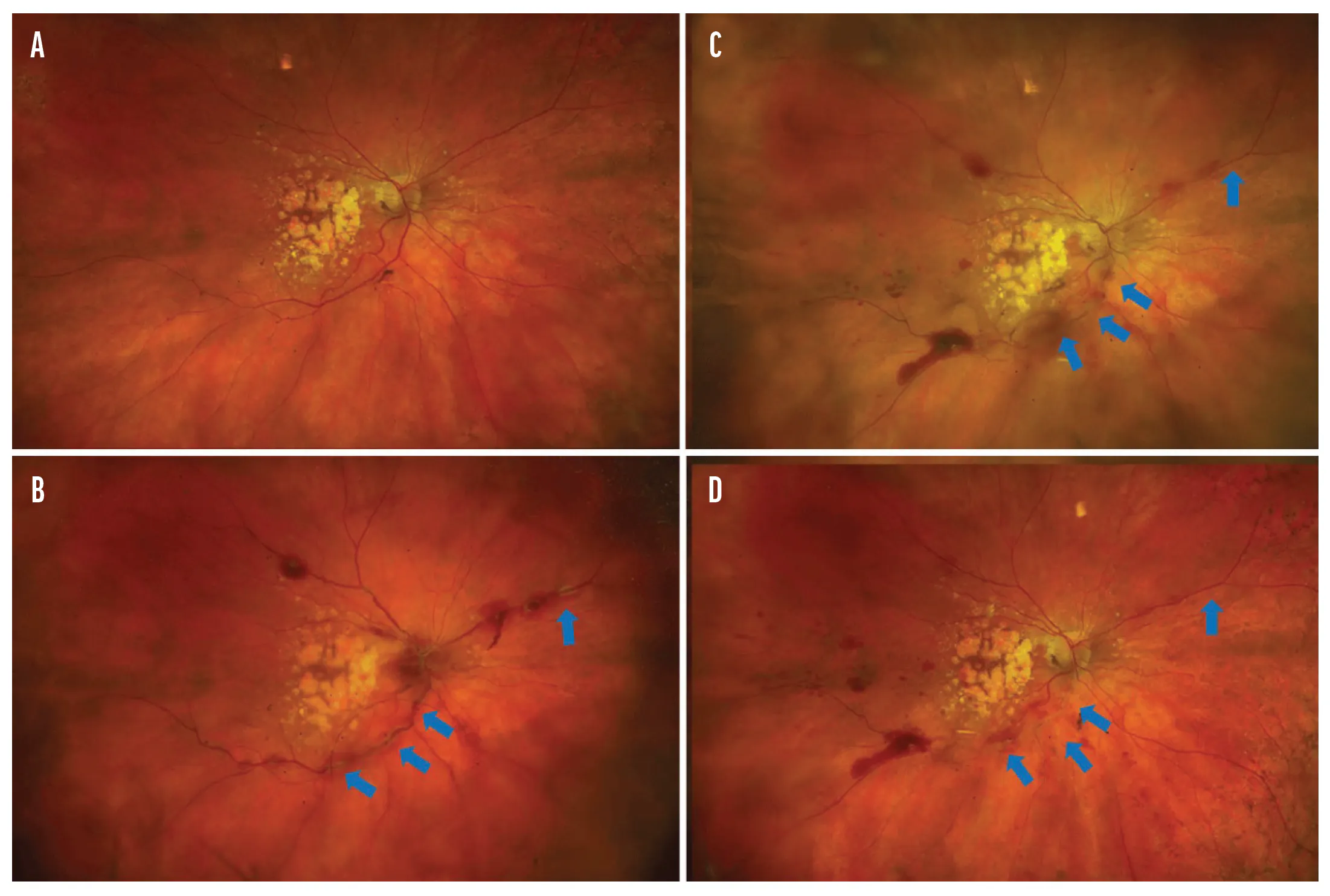
Figure 1. Baseline color fundus photograph revealed characteristics consistent with GA due to advanced dry AMD (A). After a single injection of pegcetacoplan, the patient returned to the clinic. Color fundus photograph revealed venous tortuosity (B, blue arrows) and disc congestion with some intraretinal hemorrhage. Optic disc congestion and scattered hemorrhage improved with steroids alone, before intravitreal ceftazidime injection. There was a new hemorrhage inferotemporally (C). Fundus photograph 5 weeks after pegcetacoplan injection, illustrating continued improvement (D).
Fluorescein angiography (FA) imaging at 30 seconds showed delayed transit, blockage due to retinal hemorrhage, and inferior vessel occlusion (Figure 2A); at 1 minute, further vascular congestion was depicted, as was nonperfusion in both the central and peripheral retina (Figure 2B). Optic nerve leakage was seen at 8 minutes (Figure 2C). Resolution of these observations was observed after treatment, as seen in Figures 2D-F.
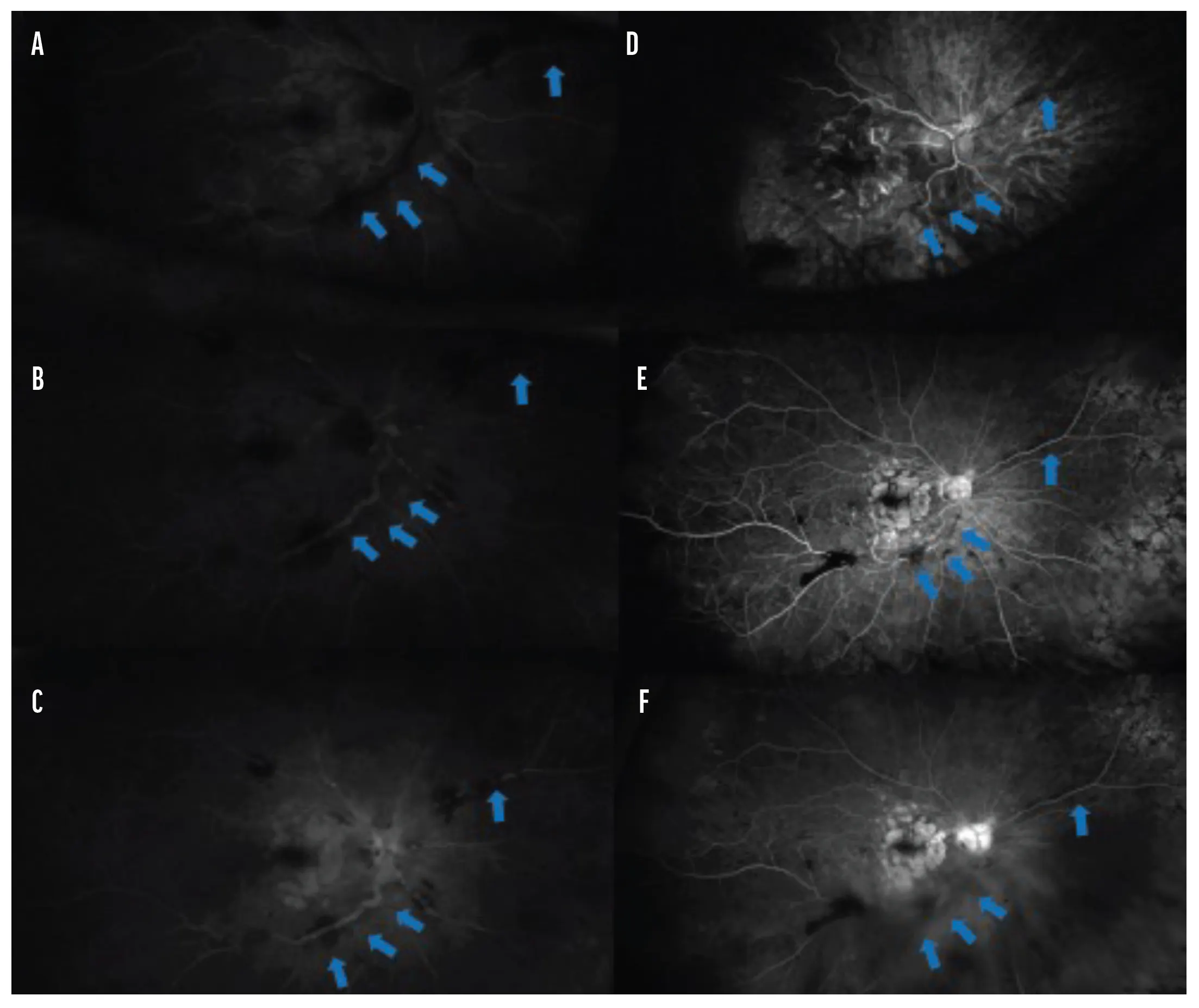
Figure 2. Fluorescein angiography (FA) revealed blocked fluorescence due to the hemorrhage at 30 seconds (A), central and peripheral nonperfusion at 1 minute (B), and optic disc leakage at 8 minutes (C). After treatment, FA showed improved arteriovenous transit time at 19 seconds (D). At 1 minute, improved central and peripheral perfusion were seen, with persistent filling defects inferiorly (E). FA at 8 minutes showed persistent late optic disc leakage (F).
OCT imaging at baseline revealed hypertransmission consistent with advanced dry AMD (Figure 3A). When the patient returned 11 days after pegcetacoplan treatment, new onset cystoid macular edema (CME) was present (Figure 3B). At this point, the patient’s VA was counting fingers, down from 20/30 before treatment.
The patient received a series of steroid, antibiotic, and pressure-lowering treatments. At presentation, intravitreal and subconjunctival dexamethasone were administered. The patient was directed to administer topical difluprednate 0.05% hourly and was also directed to administer polymyxin/neomycin hourly. Dorzolamide-timolol was directed to be administered twice daily. Prophylactic valacyclovir was prescribed, as well as a methylprednisolone dose pack (which was switched to a regimen of oral prednisone 60 mg daily the following day).
The next day, the patient reported subjective improvement of vision. Two days after presenting with complications, OCT depicted CME improvement, which corresponded with improvement to 20/200 VA (Figure 3C). The patient received a sub-Tenon’s 40 mg injection of triamcinolone.
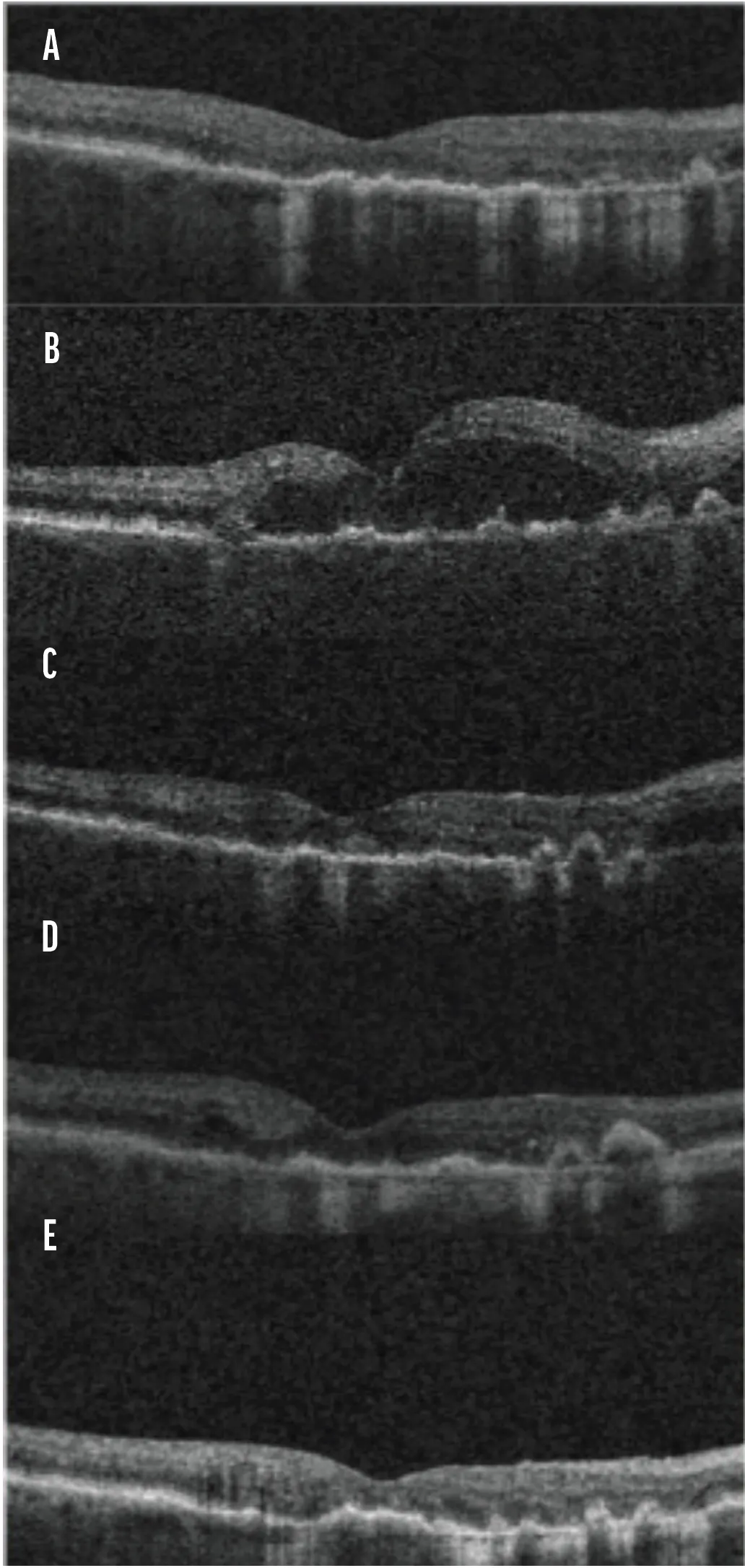
Figure 3. OCT images throughout her occlusive retinal vasculitis course. Geographic atrophy was observed at baseline (A). Upon presentation with occlusive retinal vasculitis, new cystoid macular edema (CME) was present (B). Following intravitreal and subconjunctival dexamethasone, significant improvement was observed within 2 days (C). Improvement in CME continued 2 days later following a sub-Tenon triamcinolone (D). Eight months after the onset of occlusive vasculitis, lingering CME was treated with anti-VEGF therapy (E).
Two days later (i.e., 4 days after presenting with complications), OCT showed continued improvement in CME (Figure 3D). Two days later (i.e., 6 days after presenting with complications), the patient received a 0.40 mg intravitreal injection of dexamethasone, a 4 mg subconjunctival injection of dexamethasone, a 2.25 mg intravitreal injection of ceftazidime, and 400 mg of oral moxifloxacin daily. Three days later, VA improved to 20/30, which was her VA at baseline.
Lingering recurrent CME, which was observed on OCT at 8 months post-complication, was addressed with anti-VEGF therapy (Figure 3E). VA was 20/20.
PATIENT COUNSELING INFORMATION: Advise patients that following SYFOVRE administration, they are at risk of developing endophthalmitis, retinal detachment, retinal vasculitis with or without retinal vascular occlusion and neovascular AMD. If the eye becomes red, sensitive to light, painful, or if a patient develops any change in vision such as flashing lights, blurred vision or metamorphopsia, instruct the patient to seek immediate care from an ophthalmologist. Patients may experience temporary visual disturbances associated either with the intravitreal injection of SYFOVRE or the eye examination. Advise patients not to drive or use machinery until visual function has recovered sufficiently.
ROUNDTABLE DISCUSSION
During the roundtable, Apellis posed a series of questions to the experts and what follows is a summary of their answers.
In addition to the patient counseling information in the PI, what instructions do you provide patients who receive a pegcetacoplan injection regarding potential post-injection complications?
Roger Goldberg, MD, MBA: All patients, regardless of the specific injection they receive, obtain post-injection instructions from my office. Symptoms of a post-injection complication are nonspecific, and are typically generalized complaints such as lost vision, pain, and/or new onset floaters. We remind patients that they may experience some discomfort or slight floaters after an injection, but that they should call our office if they haven’t improved within 1 to 2 days.
In my experience, patients know when something needs to be addressed. I tell them to call our office immediately if they suspect something seems off. We provide a human-monitored phone number for patients to call if they want to return for a potential post-injection visit. The easier it is to schedule an appointment, the quicker we can get patients into the clinic.
Sharon Fekrat, MD, FACS, FASRS: I tell my patients to call if they feel like they’re experiencing a complication, and I give my patients wide berth to determine what “complication” means. I always tell them to contact the office immediately if they suspect something is wrong. Providing patient counseling about what to expect following an injection helps support patient understanding and appropriate post-injection care.
I give monocular patients my cell phone number so they can contact me directly. And I tell all patients that if they can’t get ahold of someone at my office while experiencing a complication, that they should just show up to the clinic or go to the ER. The last thing I want is a patient with a complication delaying care because of a logistical scheduling issue.
Deepak Sambhara, MD, FASRS: I sometimes inject the worse-seeing eye first in a GA patient to see if any AEs occur. Patients who heavily rely on their better-seeing eye could overlook vision loss in their worse eye if the discrepancy between the two eyes is significant. Therefore, I instruct these patients to be vigilant with their worse-seeing eye to monitor potential complications after their first injection.
Walk through your workup of a patient who reports vision loss and pain several days following an injection.
Rishi P. Singh, MD: Time is of the essence in these patients. I immediately conduct an examination where I aim to detect whether the complication is infectious. If I observe hypopyon, then I immediately turn to intravitreal ceftazidime and dexamethasone after a paracentesis; if I observe occlusions, then I order a round of intravitreal steroids alone. Immediate action can allow you to treat the patient while you further investigate the source of the complication and chart a path forward. Topical agents, in my estimation, don’t adequately penetrate ocular tissue. I avoid them.
The retina specialists in the case described above by Leung used a methodology I typically reserve for patients with infectious endophthalmitis, although I do appreciate that the treatment nevertheless resulted in resolution of symptoms and vasculitis.1
Dr. Fekrat: The time of presentation may inform our diagnosis. In my clinical experience and in listening to my colleagues describe complications they’ve faced, infectious endophthalmitis typically becomes symptomatic 3 to 5 days following an injection, whereas occlusive retinal vasculitis such as that seen in the case described by Leung et al.1 typically becomes symptomatic within 9 to 11 days.
As far as the patient case seen here, I think the retina specialists’ consistent administration of steroids likely resulted in a positive outcome. It’s unclear to me, given the lack of infectious characteristics, whether the antibiotics played any role.
Dr. Sambhara: I agree that it’s unclear what role the antibiotics played in this case, but we should acknowledge that the patient’s vision improved significantly after they were administered.
Dr. Fekrat: We’ll never know for sure if this was because the steroids had time to do their job or because there was indeed an infection at play.
Dr. Goldberg: If I were to observe cells in a patient who presented with post-injection complications, I would order an ultra-widefield FA immediately, even if I didn’t observe hemorrhagic or occlusive vasculitis. You could possibly observe delayed transit in the periphery, even if it falls short of a full occlusion.
Dr. Singh: The three imaging modalities used here are OCT, FA, and CFP. These modalities are great places to start. Use of a red filter could be useful in cases where hemorrhage interferes with a clear image, and leveraging ultra-widefield technology gives us the most data possible for these important cases.
"I have recently seen patients with GA who came to me for a second or third opinion and never had a conversation with their retina specialists about GA management. These novel AI tools may open the door in the future to more conversations between patients and retina specialists.”
— DR. SINGH
How frequently would you direct a patient with retinal vasculitis to return to your clinic?
Dr. Singh: I applaud the retina specialists in this case for their close observation of the patient. I, too, would follow this patient daily until substantial improvement is realized. Daily observation allows us to catch neovascularization, hemorrhage, or a missed infection.
The patient in this case received intravitreal, sub-Tenon’s, and subconjunctival steroids. Which routes of administration do you most value when treating retinal vasculitis?
Dr. Fekrat: In my estimation, the route of administration is less important than the timing. As long as we act fast, we’re keeping the priority in focus.
Dr. Singh: Sub-Tenon’s steroid injections work well for pathologies such as uveitis, but in this case, I think sub-Tenon’s injection and subconjunctival injections duplicate the effort of an intravitreal steroid injection.
What are some factors you personally keep top of mind when considering treating a patient with pegcetacoplan?
Dr. Fekrat: Careful patient evaluation and monitoring remain important considerations in clinical practice. Balancing the potential efficacy with safety risks upfront is always important when counseling every patient. I contextualize these safety data with patients to make sure we’re making the most informed decision possible together.
Dr. Singh: I share that perspective. Also, it’s reassuring to see the increased awareness around retinal vasculitis as well as pegcetacoplan as a treatment option amongst HCPs. Based on our training, close monitoring for early signs and symptoms is critical to every management plan; retina specialists have become more cautious when monitoring patients with a history of ocular inflammation.
Dr. Sambhara: I agree with both Dr. Singh and Dr. Fekrat. Careful patient selection and assessment remain important aspects of clinical practice. Reviewing a patient’s medical and ocular history—including a history of autoimmune or inflammatory disease when relevant—helps retina specialists make informed treatment decisions and determine appropriate monitoring strategies.
"Careful patient evaluation and monitoring remain important considerations in clinical practice. Balancing the potential efficacy with safety risks upfront is always important when counseling every patient.”
— DR. FEKRAT
Dr. Goldberg: There is one group I would never dose with a pegcetacoplan injection: patients who already had an inflammatory response. I’m not comfortable challenging these patients again. That being said, I always have a conversation with every one of my GA patients that pegcetacoplan is a treatment option with the context of its risk-benefit profile.
Dr. Sambhara: Context is definitely king, and the more information we have as retina specialists, the better equipped we are to help our patients. I applaud Apellis for their vigilance with safety and commitment to the retina community with roundtable discussions such as this.
Similarly, alongside the continued characterization of retinal vasculitis over the past several years, emerging data have expanded our understanding of GA disease progression, and have articulated factors that may inform treatment decisions. As the field gains additional experience with the safety profile of pegcetacoplan injection, there has also been increasing interest in identifying imaging biomarkers and structural changes associated with disease progression, with the goal of helping clinicians better characterize which patients may be appropriate candidates for treatment and when intervention may be most meaningful.
Schmidt-Erfurth et al. have reported that, based on a deep learning–based post-hoc analysis of OCT data from sham patients in the pivotal phase 3 OAKS and DERBY trials, EZ loss precedes RPE loss.2
In a post-hoc analysis of OAKS and DERBY, researchers used a deep-learning algorithm to analyze volumetric OCT scans. The algorithm segmented EZ and inner RPE boundaries and measured EZ-RPE thickness. Researchers quantified photoreceptor loss by mapping EZ-RPE thickness values across the macula.
Patients with ≤4 µm of EZ thickness were considered to have EZ loss. Data showed that patients had less EZ loss if they were dosed with pegcetacoplan every month or every other month (EOM) compared with patients who received sham.5 In OAKS and DERBY at Year 2, sham patients lost approximately 4.47mm2 compared with 2.25mm2 and 2.41mm2 in the monthly and EOM groups, respectively.2 The study’s limitations should be noted. As it is a post-hoc analysis, there is no control for type 1 error, and the results should be interpreted with caution. This post-hoc study only included patients from OAKS and DERBY who had been examined with Spectralis OCT (Heidelberg Engineering), and did not include patients whose GA lesions expanded beyond the OCT field of view. Additionally, the AI algorithm used in the study has not been approved by the FDA. Figure 4 shows reduction in EZ loss on OCT over 24 months in the post-hoc OAKS and DERBY analysis, and Figure 5 shows reduction in RPE loss on OCT over 24 months.
Figure 6 illustrates how EZ and RPE loss areas would be depicted on OCT over time in a hypothetical patient.
These post-hoc data show that EZ:RPE loss ratios in OAKS and DERBY sham patients may predict GA progression rates.
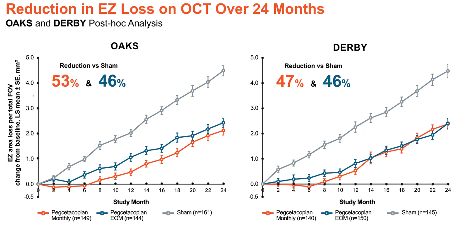
Figure 4. Reduction in EZ loss as seen on OCT over 24 months in OAKS and DERBY. The sham arm can be seen in gray.
Adapted from Schmidt-Erfurth U, et al. Ophthalmology; 2025. with permission from Elsevier.
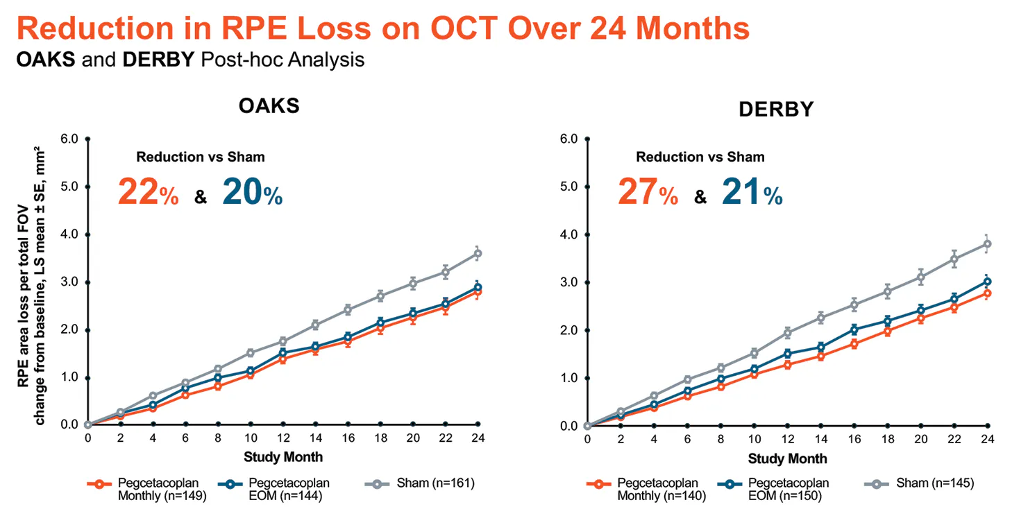
Figure 5. Reduction in RPE loss as seen on OCT over 24 months in OAKS and DERBY. The sham arm can be seen in gray.
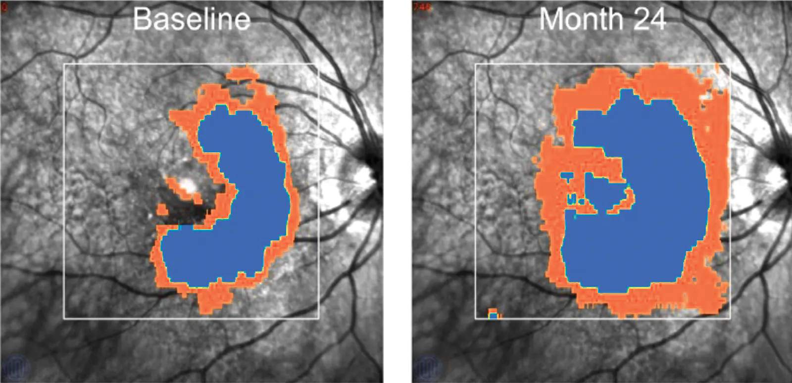
Figure 6. This illustration depicts a hypothetical patient whose EZ and RPE loss areas were measured and calculated on OCT imaging. EZ loss area is depicted in orange and RPE loss area is depicted in blue. At year 2, regions that experience only EZ loss at baseline now experience RPE loss, and regions that were untouched by pathology at baseline are experiencing EZ loss.
For illustrative purposes only. OCT-based evaluation of GA lesion progression in an untreated eye. Adapted from Schmidt-Erfurth U, et al.
Ophthalmology; 2025. with permission from Elsevier.
• The photoreceptor layer is represented on an OCT scan by the EZ layer.1
• EZ layer loss is significantly correlated to GA lesion growth in observational studies.1,2
AI = artificial intelligence; EZ = ellipsoid zone; OCT = optical coherence tomography; RPE = retinal pigment epithelium
What do you think about novel data such as that described by Schmidt-Erfurth et al?
Dr. Sambhara: It’s exciting to see research and inventive thinking in the GA space, given that we previously had no treatment. The application of AI models is particularly exciting.
The data from Schmidt-Erfurth et al. certainly make us think about GA as the inevitable ending of EZ loss, which in turn leads to RPE loss and eventual RPE atrophy. We’d all love a way to stratify risk for patients, and the team working with Schmidt-Erfurth did so by calculating the EZ:RPE loss ratio among sham eyes in OAKS and DERBY.2 These post-hoc data show that EZ:RPE loss ratio, when stratified by quartile in OAKS and DERBY sham patients, predicted GA progression rate. Those with the lowest EZ:RPE loss ratio experienced the slowest GA growth, while those with the highest EZ:RPE loss ratio experienced the most significant GA progression.2
If we had the ability to quickly assess EZ:RPE loss ratio in our clinics, then we could potentially assess patient risk based on objective metrics rather than by subjective observation.
"It’s exciting to see research and inventive thinking in the GA space, given that we previously had no treatment. The application of AI models is particularly exciting."
— DR. SAMBHARA
Dr. Goldberg: These data help illuminate our understanding of GA. I tend to use the term photoreceptor loss rather than EZ loss when discussing data such as this with patients, as I think it’s more intuitive. In fact, I even use that language when speaking with other retina specialists.
Dr. Sambhara is right that we would love to have the ability to quickly assess EZ:RPE loss ratios. The functional word is quickly. In OAKS and DERBY, study sites captured high-density scans that were time-consuming.
Dr. Fekrat: If we could find a way to assess EZ:RPE loss ratio with simpler scans and instant calculations, then we could potentially find a way to use this functionality in our clinic in the future. As it stands, retina specialists spend so much chair time with GA patients that we don’t have the time to capture imaging and calculate an EZ:RPE loss ratio. Automating this will be key.
Dr. Singh: I have recently seen patients with GA who came to me for a second or third opinion and never had a conversation with their retina specialists about GA management. These novel AI tools may open the door in the future to more conversations between patients and retina specialists.
Dr. Goldberg: We also need means of depicting the benefits of complement inhibition therapy to patients who are already undergoing treatment. Patients who elect to receive complement inhibition still lose vision and may become discouraged, thereby putting them at risk for loss to follow-up. For some patients, it would be useful to show their GA growth rate on a line chart and compare their post-treatment growth rate to their previous growth rate. Showing how the curve changes over time would help some patients understand the value of continued therapy.
Dr. Sambhara: Regardless of the specific data collected or the form it takes, we need additional tools to educate patients and retina specialists about a patient’s specific GA prognosis. The more education tools are available, the more patients can potentially understand their GA and its progression, have more informed conversations with their care team, and improve their awareness of how the disease may change over time.
1. Leung EH, Yeh S, Lampert S. Favorable outcome after pegcetacoplan (Syfovre)-associated occlusive retinal vasculitis. Ophthalmic Surg Lasers Imaging Retina. 2025;56(3):170-173.
2. Schmidt-Erfurth U, Mai J, Reiter GS, et al. Disease activity and therapeutic response to pegcetacoplan for geographic atrophy identified by deep learning-based analysis of OCT. Ophthalmology. 2025;132(2):181-193.
3. Albini R, Regillo C, Wykoff, C, et al. SYFOVRE: insights on case reports of retinal vasculitis and removal of 19-gauge filter needle. Retina Today. Insert. October 2023.
4. Data on file. Apellis Pharmaceuticals, Inc.
5. Leng T, Schneider EW, Rahimy E, et al. IRIS® Registry analysis of anti-VEGF treatment in patients with coexisting neovascular age-related macular degeneration and geographic atrophy. Oral presentation at: The Association for Research in Vision and Ophthalmology Annual Meeting; May 5-9, 2024; Seattle, Washington.
SYFOVRE® (pegcetacoplan injection) Indication & Important Safety Information
INDICATION
SYFOVRE is indicated for the treatment of geographic atrophy (GA) secondary to age-related macular degeneration (AMD)
IMPORTANT SAFETY INFORMATION
CONTRAINDICATIONS
SYFOVRE is contraindicated in patients with ocular or periocular infections, in patients with active intraocular inflammation, and in patients with hypersensitivity to pegcetacoplan or any of the excipients in SYFOVRE. Systemic hypersensitivity reactions (e.g., anaphylaxis, rash, urticaria) have occurred.
WARNINGS AND PRECAUTIONS
• Endophthalmitis and Retinal Detachments
– Intravitreal injections, including those with SYFOVRE, may be associated with endophthalmitis and retinal detachments. Proper aseptic injection technique must always be used when administering SYFOVRE to minimize the risk of endophthalmitis. Patients should be instructed to report any symptoms suggestive of endophthalmitis or retinal detachment without delay and should be managed appropriately.
• Retinal Vasculitis and/or Retinal Vascular Occlusion
– Retinal vasculitis and/or retinal vascular occlusion, typically in the presence of intraocular inflammation, have been reported with the use of SYFOVRE. Cases may occur with the first dose of SYFOVRE and may result in severe vision loss. Discontinue treatment with SYFOVRE in patients who develop these events. Patients should be instructed to report any change in vision without delay.
• Neovascular AMD
– In clinical trials, use of SYFOVRE was associated with increased rates of neovascular (wet) AMD or choroidal neovascularization (12% when administered monthly, 7% when administered every other month and 3% in the control group) by Month 24. Patients receiving SYFOVRE should be monitored for signs of neovascular AMD. In case anti-Vascular Endothelial Growth Factor (anti-VEGF) is required, it should be given separately from SYFOVRE administration.
• Intraocular Inflammation
– In clinical trials, use of SYFOVRE was associated with episodes of intraocular inflammation including: vitritis, vitreal cells, iridocyclitis, uveitis, anterior chamber cells, iritis, and anterior chamber flare. After inflammation resolves, patients may resume treatment with SYFOVRE./p>
• Increased Intraocular Pressure
– Acute increase in IOP may occur within minutes of any intravitreal injection, including with SYFOVRE. Perfusion of the optic nerve head should be monitored following the injection and managed as needed.
ADVERSE REACTIONS
• Most common adverse reactions (incidence ≥5%) are ocular discomfort, neovascular age-related macular degeneration, vitreous floaters, conjunctival hemorrhage.
Please see full Prescribing Information for more information.
For use by Apellis Medical for scientific and medical discussion only.
Do not photograph, copy or distribute.
