




KEY TAKEAWAYS
- By analyzing large volumes of data, AI could generate clinically meaningful insights to enable more informed decision making in the OR.
- AI could identify high-risk cases and flag factors such as suboptimal glycemic control, anticoagulant use, medical comorbidities, drug allergies, and mobility limitations.
- Agentic AI can act as a digital co-pilot by integrating preoperative, intraoperative, and postoperative capabilities.
Vitreoretinal surgery is a complex discipline involving intricate and highly variable pathology. Many decisions depend on dynamic changes in anatomy and tissue behavior, surgical visualization, and potential complications. As such, vitreoretinal fellows face a steep learning curve. Years of training through residency, fellowship, and independent surgical practice are required to develop the judgement needed to determine whether surgery is warranted, which intervention is most appropriate, and how urgently it should be performed.
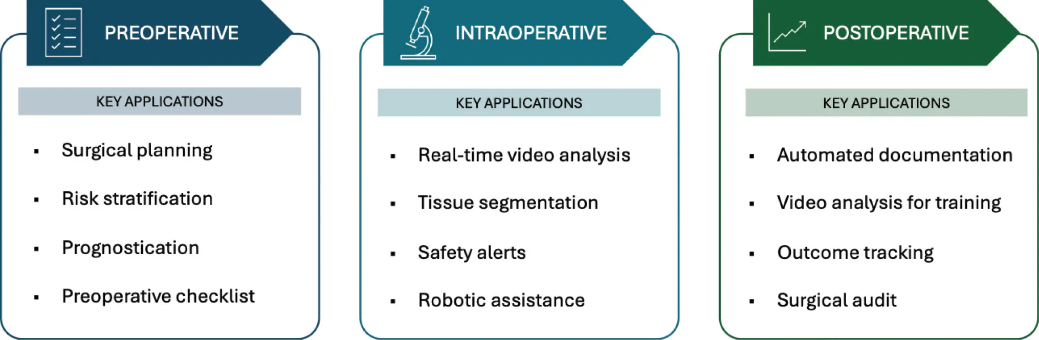
As AI tools proliferate and improve, they could advance the evolution of the retina OR. By analyzing large volumes of clinical, imaging, and procedural data, AI could generate timely, clinically meaningful insights to enable more informed decision making across preoperative, intraoperative, and postoperative phases of care (Figure).
In many ORs, digital visualization systems and intraoperative OCT have already broadened what surgeons can see and have influenced surgical decision making. These technologies can provide magnified stereoscopic visualization, digitally enhanced color contrast, and real-time imaging of retinal structures, offering invaluable guidance during surgery.
PREOPERATIVE APPLICATIONS
Every surgical case requires meticulous planning, and many decisions are made before the first trocar is inserted. This process integrates considerable data, including history, patient-specific risk factors, examination findings, and multimodal retinal imaging. For retinal detachments, for example, machine-learning models could integrate ultra-widefield imaging, OCT findings, and other clinical variables to estimate the likelihood of anatomic success. In macular surgery, AI models could help predict anatomic closure or visual outcomes using features such as macular hole dimensions, epiretinal membrane configuration, ellipsoid zone integrity, and other changes.1,2 Such predictions could improve prognostication and optimize the operative approach.
Large language models may also reduce administrative burden and improve preoperative and perioperative workflows. Studies show that these models can efficiently summarize clinic notes, extract relevant information from electronic health records (EHRs), and draft treatment plans.3,4 When appropriately integrated into EHR systems, AI could identify high-risk cases and flag factors such as suboptimal glycemic control, anticoagulant use, medical comorbidities, drug allergies, and mobility limitations. These considerations are important not only for perioperative safety but also for tailoring surgical plans to individual patient needs. AI could also generate case-specific equipment checklists based on the pathology, procedure, and surgeon’s preference, helping OR nurses and assistants support surgical workflows.
INTRAOPERATIVE APPLICATIONS
One of the most compelling potential applications of AI in the retina OR is the real-time analysis of surgical video. Early proof-of-concept studies demonstrated that deep-learning models can identify, classify, and segment instruments and ocular anatomy in vitreoretinal surgical recordings.5,6 These models may recognize the instrument being used and its position, movement, depth, and proximity to critical structures such as the macula and optic disc. Future integration with surgical visualization systems could facilitate intraoperative navigation, motion analysis, and collision avoidance.
Real-time anatomic segmentation of intraoperative OCT images could further expand these capabilities. During macular surgery, an AI-assisted system might identify residual epiretinal or internal limiting membrane, persistent traction, or a full-thickness retinal defect following release of vitreomacular traction. Such assistance could be particularly valuable during membrane dissection in proliferative vitreoretinopathy and tractional retinal detachment. Potential applications include identifying safe sites to initiate peeling, highlighting areas at risk of iatrogenic injury, distinguishing fibrovascular proliferation from the underlying retina, and monitoring retinal perfusion during fluctuations in IOP.
Robotics is another rapidly developing field in ophthalmology, as it can eliminate a surgeon’s tremor and enable instrument control with micrometer-level precision. These capabilities may be particularly valuable for maneuvers requiring extreme precision, such as subretinal injection, retinal vein cannulation, and targeted delivery of gene or cell therapies.7-10 AI can potentially enhance these platforms by incorporating preoperative imaging for anatomic localization and establishing virtual safety boundaries to limit excessive instrument movement and improve instrument stability.
POSTOPERATIVE APPLICATIONS
AI has the potential to make surgical documentation more accurate and efficient. An AI assistant could, for example, generate a draft operative note and suggest procedural codes, with surgeon review and approval.11 In an AI-integrated health system, surgical audit can be done more effectively. Intraoperative events and procedural variables can be correlated with postoperative outcomes and patient satisfaction, supporting complication surveillance, workflow optimization, and institutional quality improvement.
Vitreoretinal surgery is particularly difficult to teach because many aspects of surgical performance are difficult to assess objectively. In cataract surgery, deep-learning models were able to localize key ocular structures and instruments with high accuracy, enabling the derivation of metrics for evaluating surgical skill.12 Similar approaches can be replicated and applied to vitreoretinal surgical videos. Within each surgical phase, AI can assess instrument trajectories, tremor, proximity to critical structures, ocular centration, image focus, procedural efficiency, and time spent on key steps. For trainees, this can provide objective, actionable feedback that complements traditional assessments and learning through increasing surgical volume.
FROM ASSISTANT TO AGENT
Over the years, AI has evolved from rule-based systems into machine learning, deep learning, generative, multimodal, and now agentic systems. Agentic AI does more than generate an isolated prediction; it supports an entire clinical workflow. In the retina OR, it can act as a digital co-pilot by integrating preoperative, intraoperative, and postoperative capabilities. The digital co-pilot augments but cannot replace a surgeon’s judgment, operative decisions, and responsibility.
AUGMENT, NOT REPLACE
The retina OR of the near future is likely to become increasingly digital and interconnected. AI will play an expanding role in enhancing surgical planning, intraoperative guidance, training, documentation, and postoperative evaluation. These technologies should be thoughtfully integrated into clinical workflows, while carefully considering their limitations, risks, and need for appropriate oversight. The surgeon’s ultimate responsibility is to align surgical goals with each patient’s needs. AI may support and inform these decisions, but it cannot assume responsibility for them.
For now, AI in the retina OR is best understood as an emerging digital co-pilot. Its promise lies not in autonomous surgery but in augmented surgery: more structured planning, improved visualization, greater surgical efficiency and safety, and more effective postoperative learning. The goal is not to make surgery less human, but to provide surgeons with better information the moment it’s needed.
AI disclosure: GPT-5.6 Sol (OpenAI) was used solely for grammatical refinement. The authors reviewed and approved the final manuscript and take full responsibility for its content.
1. Poh SSJ, Sia JT, Yip MYT, et al. Artificial intelligence, digital imaging, and robotics technologies for surgical vitreoretinal diseases. Ophthalmol Retina. 2024;8(7):633-645. doi.org/10.1016/j.oret.2024.01.018
2. Bernardi E, Ferro Desideri L, Shah N, et al. Artificial intelligence in vitreoretinal surgery: a systematic review of current applications and future directions. Ophthalmol Ther. 2026;15(4):1233-1250. doi.org/10.1007/s40123-026-01347-8
3. Carlà MM, Gambini G, Baldascino A, et al. Exploring AI-chatbots’ capability to suggest surgical planning in ophthalmology: ChatGPT versus Google Gemini analysis of retinal detachment cases. Br J Ophthalmol. 2024;108(10):1457-1469. doi.org/10.1136/bjo-2023-325143
4. Cohen L, Gupta AR, Patel P, Gill GS, Bains H, Gupta S. The role of large language models in ophthalmology: a review of current applications, performance, and future directions. Cureus. 17(11):e97374. doi.org/10.7759/cureus.97374
5. Baldi PF, Abdelkarim S, Liu J, To JK, Ibarra MD, Browne AW. Vitreoretinal surgical instrument tracking in three dimensions using deep learning. Transl Vis Sci Technol. 2023;12(1):20. doi.org/10.1167/tvst.12.1.20
6. Nespolo RG, Yi D, Cole E, Wang D, Warren A, Leiderman YI. Feature tracking and segmentation in real time via deep learning in vitreoretinal surgery-a platform for artificial intelligence-mediated surgical guidance. Ophthalmol Retina. 2023;7(3):236-242. doi.org/10.1016/j.oret.2022.10.002
7. Cehajic-Kapetanovic J, Xue K, Edwards TL, et al. First-in-human robot-assisted subretinal drug delivery under local anesthesia. Am J Ophthalmol. 2022;237:104-113. doi.org/10.1016/j.ajo.2021.11.011
8. Edwards TL, Xue K, Meenink HCM, et al. First-in-human study of the safety and viability of intraocular robotic surgery. Nat Biomed Eng. 2018;2:649-656. doi.org/10.1038/s41551-018-0248-4
9. Faridpooya K, van Romunde SHM, Manning SS, et al. Randomised controlled trial on robot-assisted versus manual surgery for pucker peeling. Clin Experiment Ophthalmol. 2022;50(9):1057-1064. doi.org/10.1111/ceo.14174
10. Willekens K, Gijbels A, Smits J, et al. Phase I trial on robot assisted retinal vein cannulation with ocriplasmin infusion for central retinal vein occlusion. Acta Ophthalmol (Co-penh). 2021;99(1):90-96. doi.org/10.1111/aos.14480
11. Lee YM, Bacchi S, Sia D, Casson RJ, Chan W. Optimising vitrectomy operation note coding with machine learning. Clin Experiment Ophthalmol. 2023;51(6):577-584. doi.org/10.1111/ceo.14257
12. Yeh HH, Jain AM, Fox O, Sebov K, Wang SY. PhacoTrainer: Deep learning for cataract surgical videos to track surgical tools. Transl Vis Sci Technol. 2023;12(3):23. doi.org/10.1167/tvst.12.3.23


