



A 73-year-old woman with intermediate AMD presented with a subfoveal drusenoid pigment epithelial detachment (PED) with a hypertransmission defect on OCT and a VA of 20/40 OS (Figure 1). At a 6-month follow-up appointment, the drusenoid PED was collapsing, and her VA had worsened to 20/80 OS.
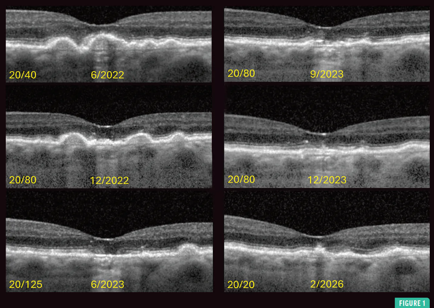
Six months later, retinal pigment epithelium (RPE) and ellipsoid zone (EZ) atrophy of > 250 µm was noted with intact overlying outer retina, and a VA of 20/125 OS. However, 6 months later, the RPE had reformed, the EZ was recovering, the hypertransmission defect had resolved, and VA improved to 20/80 OS. Fundus imaging demonstrated early atrophy that seemed to resolve over time (Figure 2).
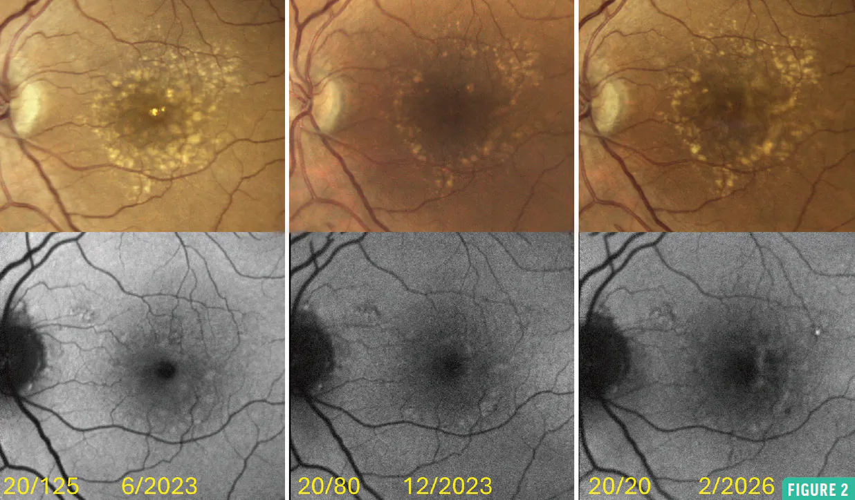
Her VA gradually improved to 20/20 OS 3 years later with overall improvement in OCT layers and no choroidal neovascularization on OCT angiography. Of note, the patient did not receive AMD treatment, nor did she experience any change in her medical history. It’s worth noting that although anticomplement therapy may have been reasonable, the patient did not yet met the criteria for geographic atrophy.
THE ROLE OF THE OUTER RETINAL LAYER
The classification of atrophy meetings (CAM) criteria describe precursor stages for inevitable geographic atrophy development.1,2 The present case, in which the outer retinal layer appeared unaffected while the RPE and EZ were disrupted, did not fit into the CAM classification. The maintenance of the outer retinal layer implies that the photoreceptor cells were possibly still viable. While further study is necessary, this case suggests that outer retinal layer thinning may be key for developing atrophy.
1. Sadda SR, Guymer R, Holz FG, et al. Consensus definition for atrophy associated with age-related macular degeneration on OCT: Classification of Atrophy Report 3. Ophthalmology. 2018;125(4):537-548. doi.org/10.1016/j.ophtha.2017.09.028
2. Corradetti G, Corvi F, Nittala MG, et al. Natural history of incomplete retinal pigment epithelial and outer retinal atrophy in age-related macular degeneration. Can J Ophthalmol. 2021;56(5):325-334. doi.org/10.1016/j.jcjo.2021.01.005


