


KEY TAKEAWAYS
- The author presents a case of a 78-year-old woman who was referred to the clinic for revision surgery after experiencing worsening visual acuity and enlargement of her central scotoma following idiopathic macular hole surgery performed at another institution, which included implantation of a human amniotic membrane.
- An initial revision surgery using fluid-air exchange followed by long-acting gas tamponade was unsuccessful, prompting the authors to consider autologous retinal transplantation, resulting in anatomic closure and subjective reduction of the visual scotoma.
- The author theorizes that, in this case, the submacular embedded graft failed to achieve hole closure and appeared to interfere with normal retina-retinal pigment epithelium interaction.
A 78-year-old woman underwent combined 25-gauge phacoemulsification and pars plana vitrectomy with gas tamponade in a hospital setting for treatment of an idiopathic macular hole. Following surgery, she experienced worsening visual acuity and enlargement of her central scotoma. She was referred to our clinic for further evaluation.
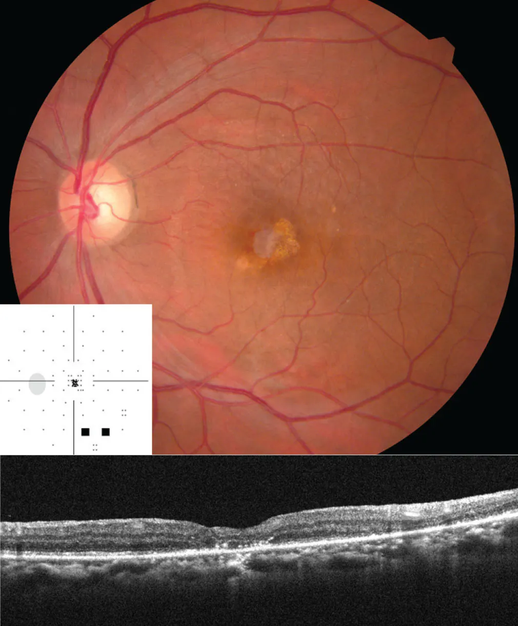
At presentation, her BCVA was 20/25 OD and 20/400 OS. Fundus examination revealed a sheet-like material located beneath the macula (Figure 1). OCT demonstrated that the edge of the sheet was embedded within the inner retinal layers, while the macular hole remained open above the implant.
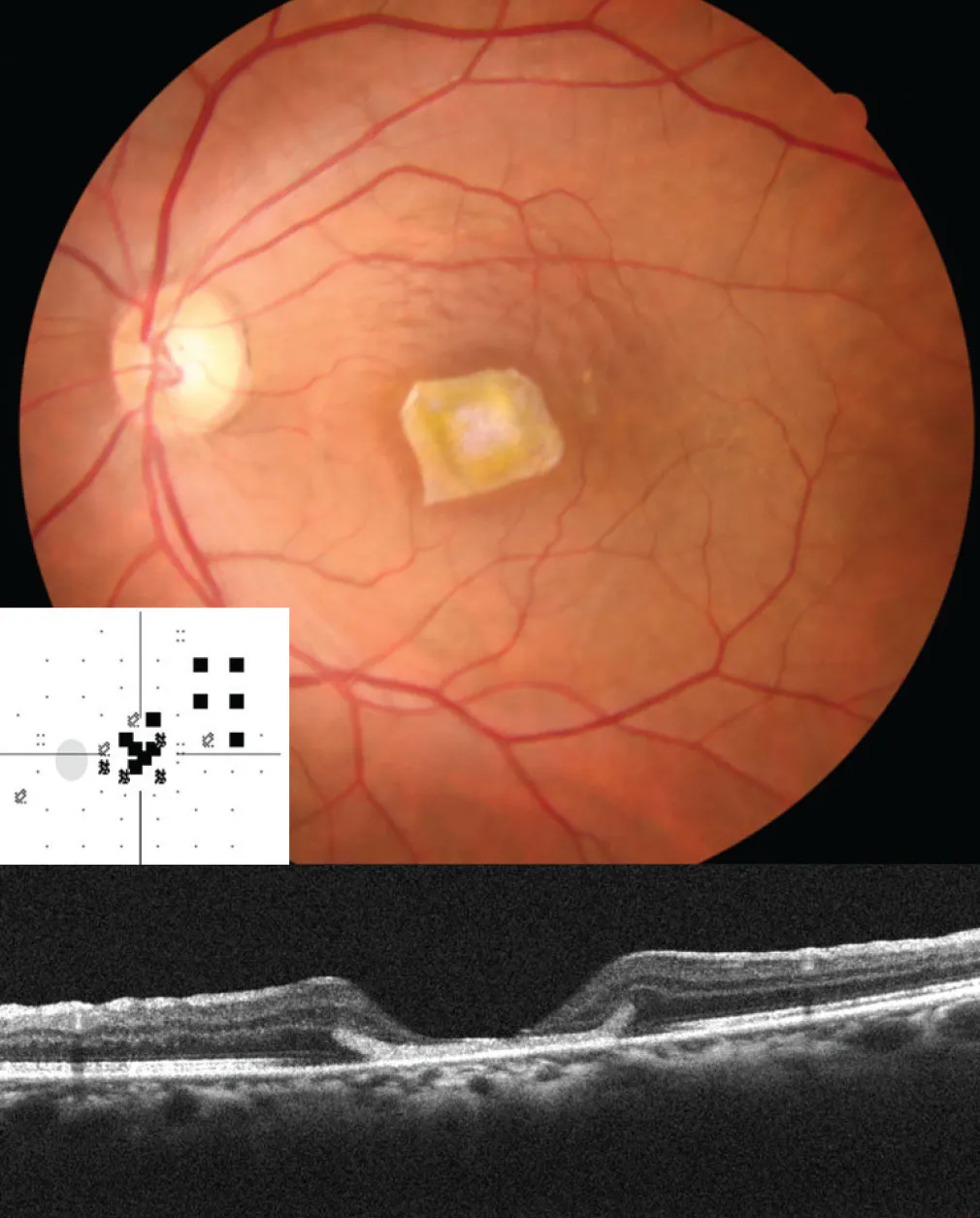
Figure 1. Fundus photography shows a HAM underneath the macula with the edge embedded within the inner retinal layers, as shown on OCT. The macular hole remained open.
SECOND SURGERY
The patient was unaware of the nature of the implanted material. Based on its appearance, location, and the presumed surgical technique, the graft was thought to be a dehydrated or cryopreserved human amniotic membrane (HAM) implanted into the submacular space during her previous surgery.1 Although the graft had been accurately positioned beneath the retina, the macular hole remained open, and the patient’s visual symptoms progressively worsened.
Because the patient strongly requested removal of the HAM, revision surgery was performed after a detailed discussion of the potential risks and benefits. Intraoperatively, extensive internal limiting membrane (ILM) removal from the previous surgery was confirmed. A localized retinal detachment was created around the graft via subretinal injection of balanced salt solution. Additional hydraulic dissection was then performed to separate the adhesion between the macular hole edge and the implanted membrane, allowing successful mobilization of the graft, which was subsequently excised with intraocular scissors and removed using forceps (Video 1).
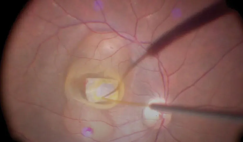
Video 1. Surgery to Remove Previous Human Amniotic Membrane and Repair Refractory Macular Hole.
As the macular hole was relatively small, we anticipated that HAM removal combined with subretinal fluid application would reproduce the principle of the subretinal fluid application technique for refractory macular holes described by Meyer et al.2 Therefore, fluid-air exchange followed by long-acting gas tamponade was performed without additional tissue transplantation.
Despite our initial optimism, the macular hole remained open after gas absorption (Figure 2). Fundus autofluorescence demonstrated extensive retinal pigment epithelium (RPE) alteration surrounding the hole. We speculate that degeneration of the underlying RPE impaired retinal migration and prevented spontaneous hole closure despite successful hydrodissection.
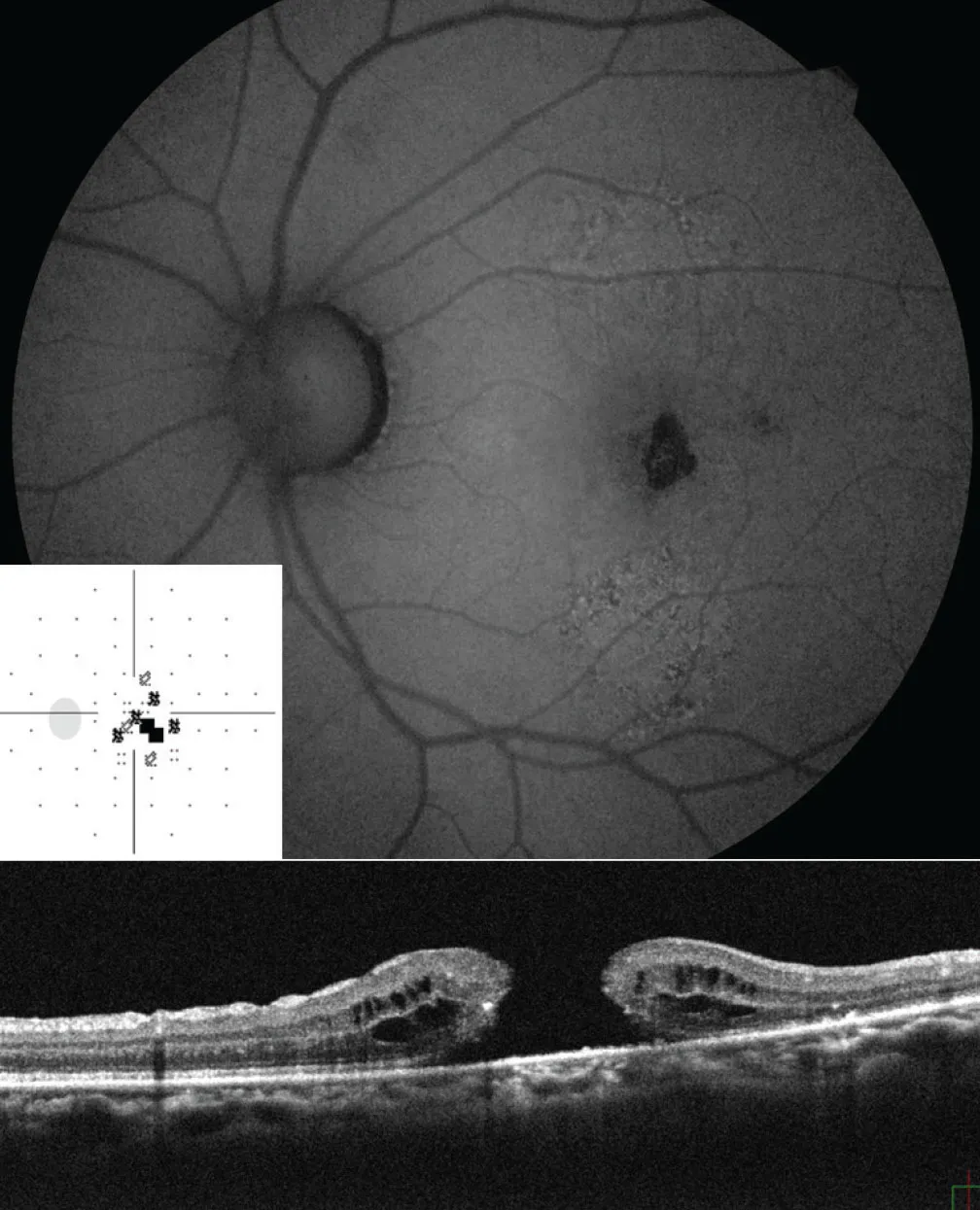
Figure 2. After the second surgery, during which the subretinal fluid application technique was used, the macular hole had yet to close.
CHANGING COURSE
Additional treatment options were subsequently considered. Because extensive ILM removal had already been performed during the primary surgery, both the inverted ILM flap technique and free ILM transplantation were not feasible. Furthermore, the hydrodissection effect achieved during the revision surgery was insufficient to close the hole.
Therefore, autologous retinal transplantation (ART), originally described in 2016 by Grewal et al, was performed.3 The retinal graft successfully sealed the defect and achieved anatomic closure of the macular hole (Video 2; Figure 3). Her VA remained unchanged at 20/400, which was not unexpected given the chronic retinal damage, but she reported a marked reduction in the size of the central scotoma and was satisfied with the functional outcome.
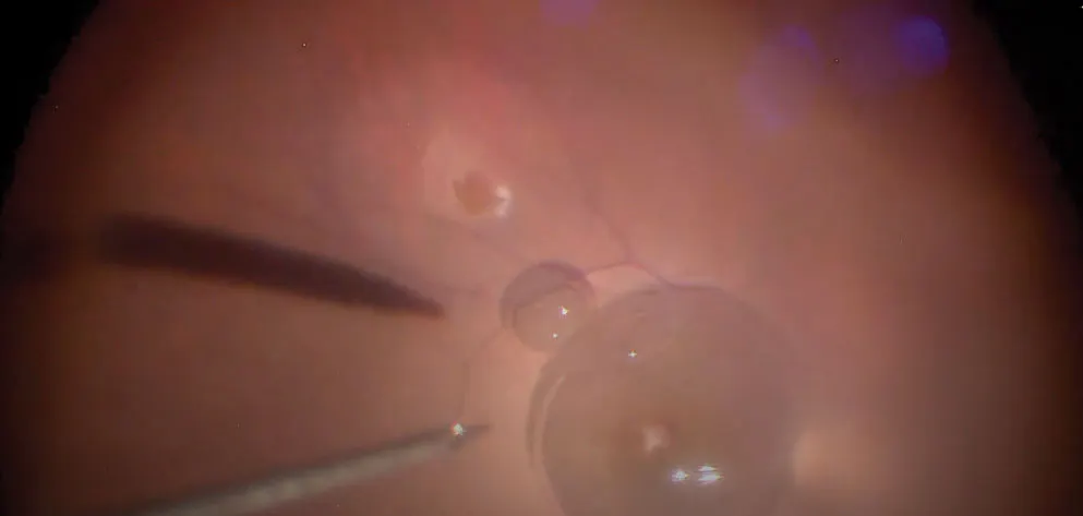
Video 2. Autologous Retinal Transplantation for Macular Hole Repair.
LESSONS LEARNED
HAM transplantation has recently emerged as a potential option for refractory macular holes, serving as a scaffold for retinal tissue proliferation and migration.4,5 In this case, however, the submacular embedded graft failed to achieve hole closure and appeared to interfere with normal retina-RPE interaction. Removal of the graft was technically feasible using controlled subretinal fluid application and hydraulic dissection. Nevertheless, hydrodissection alone was insufficient to achieve closure, likely because of irreversible RPE dysfunction surrounding the hole.
This case underscores the importance of appropriate graft sizing and selection of the HAM type in submacular amniotic membrane implantation. When conventional techniques fail and ILM is unavailable, ART remains a valuable salvage procedure capable of achieving anatomic closure and improving subjective visual function.
1. Caporossi T, De Angelis L, Pacini B, et al. Human amniotic membrane plug to promote failed macular hole closure. Sci Rep. 2020;10:18264. doi.org/10.1038/s41598-020-75292-2
2. Meyer CH, Szurman P, Haritoglou C, et al. Application of subretinal fluid to close refractory full-thickness macular holes: treatment strategies and primary outcome: APOSTEL study. Graefes Arch Clin Exp Ophthalmol. 2020;25:2151-2161. doi.org/10.1007/s00417-020-04735-3
3. Grewal DS, Mahmoud TH. Autologous neurosensory retinal free flap for closure of refractory myopic macular holes. JAMA Ophthalmol. 2016;134(2):229-230. doi.org/10.1001/jamaophthalmol.2015.5237
4. Rizzo S, Caporossi T, Tartaro R, et al. A human amniotic membrane plug to promote retinal breaks repair and recurrent macular hole closure. Retina. 2019;39 Suppl 1:S95-S103. doi.org/10.1097/IAE.0000000000002320
5. Zhang H, Wang J, Chen Y, et al. Human amniotic membrane graft for refractory macular hole: a systematic review and meta-analysis. J Fr Ophtalmol. 2023;46:226-235. doi.org/10.1016/j.jfo.2022.07.001


