




KEY TAKEAWAYS
- In October 2025, the AAO updated its 2016 guidelines on screening for hydroxychloroquine toxicity, now recommending SD-OCT and widefield fundus autofluorescence as primary screening tests.
- Visual fields, once considered a primary screening tool, is now a confirmatory test, as is multifocal electroretinogram.
- Novel imaging technologies are under investigation for their potential utility in hydroxychloroquine screening, such as adaptive optics and fluorescence lifetime imaging ophthalmoscopy.
Hydroxychloroquine is used to treat systemic lupus erythematosus, rheumatoid arthritis, sarcoidosis, and other autoinflammatory and dermatologic conditions.1 In addition, researchers are investigating its potential use as an adjunct cancer therapy.1 The recommended dose is < 5 mg/kg/day based on real body weight, which originates from a study by Melles et al showing that the likelihood of toxicity was < 1% within 5 years and < 2% after 10 years of treatment with hydroxychloroquine.2 High doses, long duration, and risk factors such as age, renal disease, tamoxifen use, or underlying macular disease can cause retinal toxicity with the use of hydroxychloroquine, with damage first occurring in the photoreceptors and outer retina, and eventually the retinal pigment epithelium (RPE).3 The classic pattern of hydroxychloroquine toxicity is the development of bull’s-eye maculopathy caused by parafoveal RPE depigmentation (Figure 1).4
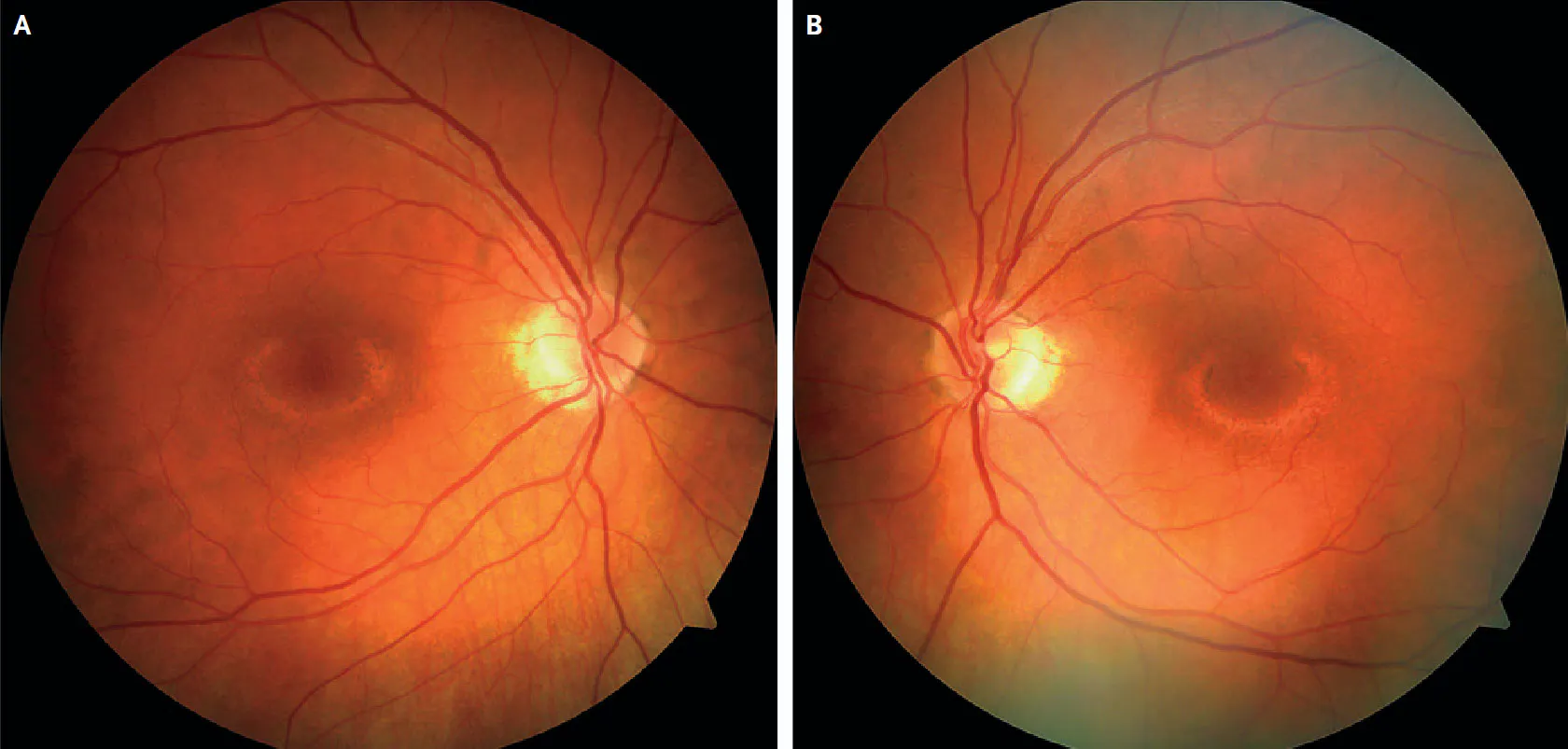
Figure 1. Bull’s-eye maculopathy from hydroxychloroquine use. Reprinted with permission from Modi et al.4
However, this is a late manifestation of toxicity, and the goal of the AAO’s Screening Guidelines for Hydroxychloroquine Retinopathy is to recognize early evidence of toxicity to prevent vision loss.5
In October 2025, the AAO updated its 2016 guidelines on screening for hydroxychloroquine toxicity.5,6 In this article, we highlight the changes you need to be aware of.
PRIMARY SCREENING TESTS
The previous 2016 AAO guidelines recommended automated visual fields and spectral-domain OCT (SD-OCT) as the primary tests for routine screening; multifocal electroretinography (mfERG) and fundus autofluorescence (FAF) were recommended as adjunct tests to obtain as needed.6
The 2025 guidelines still recommend SD-OCT as one of the primary tests for screening, but they no longer recommend visual fields as a primary screening modality due to its subjectivity and wide variability.5 Instead, they now recommend widefield FAF in addition to SD-OCT as primary screening tests.5 SD-OCT can show characteristic retinal changes from hydroxychloroquine damage, including initial outer retinal thinning. It can also document later parafoveal patterns of damage most often seen in European patients, and pericentral patterns of disease most often seen in East Asian patients.7 For detecting pericentral patterns of damage, widefield FAF is particularly helpful for imaging beyond the central macula. Early signs of toxicity on FAF are represented by areas of increased autofluorescence, while more severe signs of toxicity are visualized as dark areas on FAF due to missing photoreceptors and damaged RPE (Figure 2).8
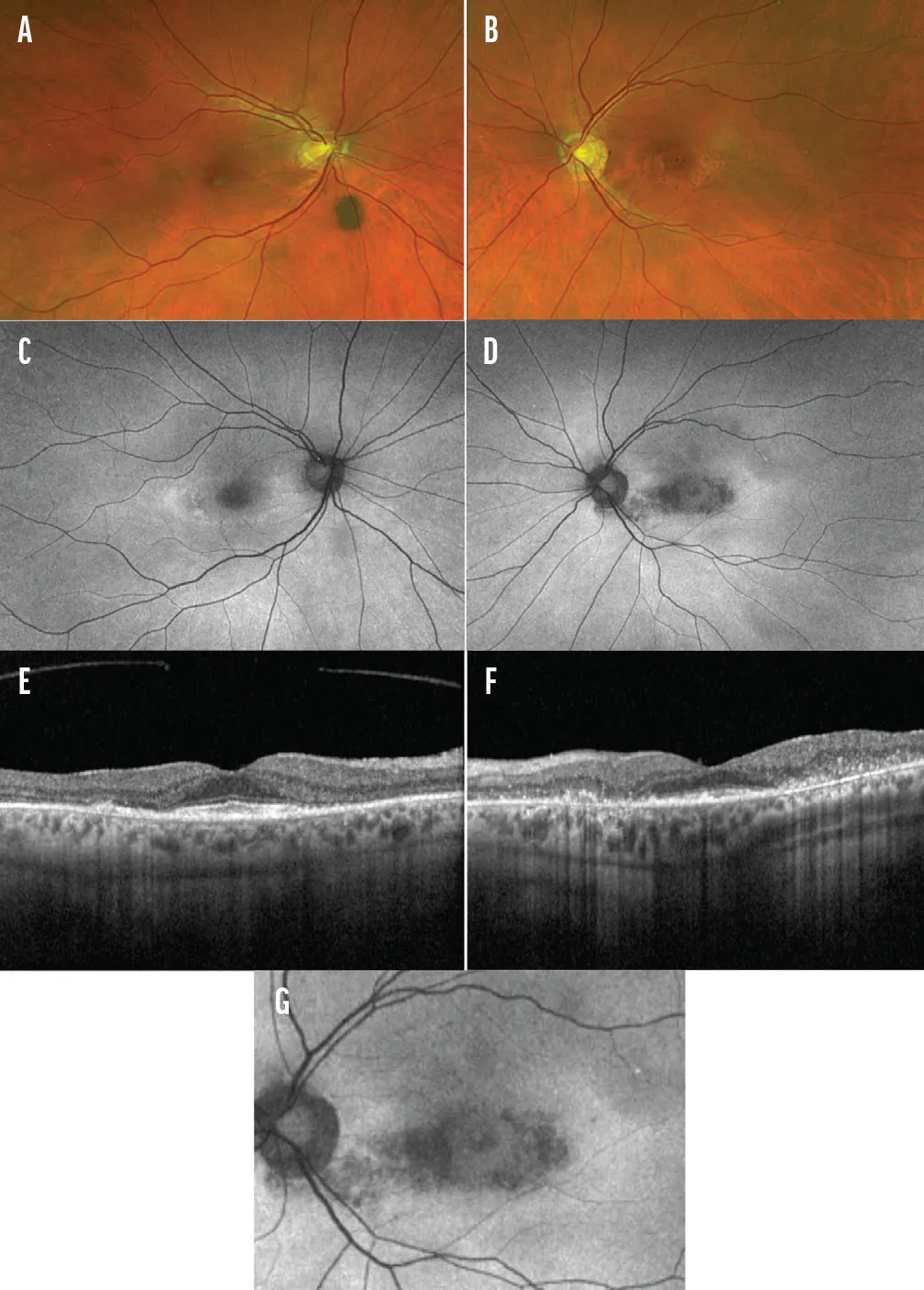
Figure 2. Multimodal imaging of a patient with hydroxychloroquine toxicity demonstrating asymmetric progression. Color fundus photography demonstrates subtle pericentral pigmentary alterations in the right eye (A) and advanced, confluent pericentral macular depigmentation in the left eye (B). FAF reveals a classic hyperautofluorescent pericentral ring, signaling outer retinal stress and lipofuscin accumulation in the right eye (C) and a well-defined zone of patchy hypoautofluorescence secondary to RPE atrophy, bordered by a hyperautofluorescent transition zone in the left eye (D). SD-OCT of the right eye shows localized parafoveal ellipsoid zone disruption and external limiting membrane with foveal sparing (“flying saucer” sign) in the right eye (E) and severe, widespread outer retinal excavation, extensive ellipsoid zone/external limiting membrane loss, and thinning of the outer nuclear layer in the left eye (F). A magnified FAF image of the left macula highlights the geographic boundaries of RPE atrophy and the surrounding hyperautofluorescent ring of active photoreceptor degeneration (G). Images courtesy of Anjali Shah, MD, University of Michigan.
SECONDARY SCREENING TESTS
Visual fields are now recommended as a confirmatory test that can be performed as needed and should include both central and peripheral testing (eg, 24-2C test). Another secondary test is mfERG, which examines responsiveness of the retina to light and can reveal ERG depression in parafoveal or extramacular areas. Tests not recommended for annual screening purposes include fundus examination and photography (visible damage on examination/photography implies an advanced stage of RPE damage), OCT angiography (vascular changes on OCT angiography suggest severe macular damage), and color vision testing (not sensitive or specific for hydroxychloroquine toxicity).5
SCREENING TIMELINE RECOMMENDATIONS
The 2016 guidelines recommended a baseline fundus examination to rule out any underlying disease or preexisting retinal damage before starting hydroxychloroquine and only obtaining SD-OCT and visual fields if maculopathy is present.6
The 2025 guidelines recommend SD-OCT and widefield FAF in addition to the fundus examination during the baseline screening for comparison with later imaging.5 The new guidelines also recommend annual screening with SD-OCT and widefield FAF upon initiation of hydroxychloroquine. However, during the first 5 years of hydroxychloroquine use, annual screening can be deferred if there are no significant risk factors for retinopathy, including renal disease, concurrent tamoxifen use, and starting hydroxychloroquine after 45 years of age.9
NEW AI APPLICATIONS IN SCREENING EFFORTS
AI has shown some promise in recognizing signs of hydroxychloroquine retinopathy on various imaging modalities. For example, Kulyabin et al studied the use of AI algorithms to interpret raw multifocal electoretinogram traces to detect retinal damage from hydroxychloroquine.1 Kalra et al used an automated machine-learning model to detect hydroxychloroquine retinopathy and predict the development of retinal toxicity using spectral-domain OCT measurements such as partial ellipsoid zone (EZ) attenuation, in addition to the duration and dose of hydroxychloroquine therapy.2 Finally, De Silva et al developed a deep-learning algorithm to automate the process of detecting and quantifying EZ loss on spectral-domain OCT images, noting that the algorithm could quantify EZ loss in patients with varying levels of toxicity, from those with only subtle signs of toxicity to those with severe toxicity and vision loss.3 Overall, AI is promising, but further research is needed to validate and standardize AI models for use in clinical practice.
1. Kulyabin M, Kremers J, Holbach V, Maier A, Huchzermeyer C. Artificial intelligence for detection of retinal toxicity in chloroquine and hydroxychloroquine therapy using multifocal electroretinogram waveforms. Sci Rep. 2024;14(1):24853. doi.org/10.1038/s41598-024-76943-4
2. Kalra G, Talcott KE, Kaiser S, Ugwuegbu O, Hu M, Srivastava SK, Ehlers JP. Machine learning–based automated detection of hydroxychloroquine toxicity and prediction of future toxicity using higher-order OCT biomarkers. Ophthalmol Retina. 2022;6(12):1241-1252. doi.org/10.1016/j.oret.2022.05.031
3. De Silva T, Jayakar G, Grisso P, Hotaling N, Chew EY, Cukras CA. Deep learning-based automatic detection of ellipsoid zone loss in spectral-domain OCT for hydroxychloroquine retinal toxicity screening. Ophthalmol Sci. 2021;1(4):100060. doi.org/10.1016/j.xops.2021.100060
NEW DIAGNOSTIC TECHNOLOGIES
Novel imaging technologies are under investigation for their potential utility in hydroxychloroquine screening, such as adaptive optics (AO), which allows for the visualization of retinal cells in high resolution. Jorge et al compared individuals with no history of hydroxychloroquine use to individuals on hydroxychloroquine who had no evidence of retinal toxicity on SD-OCT, visual fields, or mfERG.10 The researchers found that the group taking hydroxychloroquine had lower density and larger spacing of cones on AO imaging, which highlights that AO may have the potential to detect hydroxychloroquine retinal toxicity before other imaging modalities.10
Another area of research is in advanced autofluorescence techniques, including fluorescence lifetime imaging ophthalmoscopy (FLIO), which measures how long retinal fluorophores glow after excitation by laser pulses. While Solberg et al did not find a statistically significant difference in fluorescence lifetimes between healthy controls and patients on hydroxychloroquine therapy with no clinical retinopathy,11 Sauer et al found that clinically normal patients on hydroxychloroquine therapy had prolonged fluorescence lifetimes compared with healthy individuals not on hydroxychloroquine, highlighting that FLIO may be able to detect hydroxychloroquine toxicity at its beginning stages.12
SCREEN EARLY, SCREEN OFTEN
As a key long-term treatment option for many systemic conditions, hydroxychloroquine is a relatively common finding on patients’ medication lists. Thus, clinicians must be prepared to screen carefully to ensure they catch retinal toxicity early and avoid vision loss whenever possible.
1. Plantone D, Koudriavtseva T. Current and future use of chloroquine and hydroxychloroquine in infectious, immune, neoplastic, and neurological diseases: a mini-review. Clin Drug Investig. 2018;38(8):653-671. doi.org/10.1007/s40261-018-0656-y
2. Melles RB, Marmor MF. The risk of toxic retinopathy in patients on long-term hydroxychloroquine therapy. JAMA Ophthalmol. 2014;132(12):1453-1460. doi.org/10.1001/jamaophthalmol.2014.3459
3. Marmor MF. Comparison of screening procedures in hydroxychloroquine toxicity. Arch Ophthalmol. 2012;130(4):461-469. doi.org/10.1001/archophthalmol.2011.371
4. Modi YS, Singh RP. Bull’s-eye maculopathy associated with hydroxychloroquine. N Engl J Med. 2019;380(17):1656. doi.org/10.1056/NEJMicm1412167
5. Marmor MF, Ahn SJ, Ehlers JP, Melles RB, Mieler WF, Sarraf D, Yusuf IH. Special AAO Report: Recommendations on screening for hydroxychloroquine retinopathy (2025 revision). Ophthalmology. 2026;133(4):439-450. doi.org/10.1016/j.ophtha.2025.11.001
6. Marmor MF, Kellner U, Lai TYY, Melles RB, Mieler WF, American Academy of Ophthalmology. Recommendations on screening for chloroquine and hydroxychloroquine retinopathy (2016 revision). Ophthalmology. 2016;123(6):1386-1394. doi.org/10.1016/j.ophtha.2016.01.058
7. Melles RB, Marmor MF. Pericentral retinopathy and racial differences in hydroxychloroquine toxicity. Ophthalmology. 2015;122(1):110-116. doi.org/10.1016/j.ophtha.2014.07.018
8. Kellner U, Renner AB, Tillack H. Fundus autofluorescence and mfERG for early detection of retinal alterations in patients using chloroquine/hydroxychloroquine. Invest Ophthalmol Vis Sci. 2006;47(8):3531-3538. doi.org/10.1167/iovs.05-1290
9. Jorge AM, Melles RB, Marmor MF, Zhou B, Zhang Y, Choi HK. Risk factors for hydroxychloroquine retinopathy and its subtypes. JAMA Netw Open. 2024;7(5):e2410677. doi.org/10.1001/jamanetworkopen.2024.10677
10. Braga JPR, Lucena MM, Rodrigues MW, et al. Adaptive optics cone arrangement in hydroxychloroquine users without signs of retinal toxicity based on current screening guidelines: a case-control study. Graefes Arch Clin Exp Ophthalmol. 2025;263(5):1279-1287. doi.org/10.1007/s00417-025-06772-2
11. Solberg Y, Dysli C, Möller B, Wolf S, Zinkernagel MS. Fluorescence lifetimes in patients with hydroxychloroquine retinopathy. Invest Ophthalmol Vis Sci. 2019;60(6):2165-2172. doi.org/10.1167/iovs.18-26079
12. Sauer L, Calvo CM, Vitale AS, Henrie N, Milliken CM, Bernstein PS. Imaging of hydroxychloroquine toxicity with fluorescence lifetime imaging ophthalmoscopy. Ophthalmol Retina. 2019;3(10):814-825. doi.org/10.1016/j.oret.2019.04.025


