





KEY TAKEAWAYS
- The authors describe a case of atypical herpes simplex virus-2 associated bilateral acute retinal necrosis (BARN) in a monocular immunocompetent individual 30 years after involvement of the first eye.
- Antiviral therapy significantly reduces the risk of bilateral involvement with ARN, with one study suggesting 87.1% of patients treated with acyclovir had fellow eyes that remained disease-free, compared with 30.4% of untreated patients.
- The development of a rash nonresponsive to systemic steroids or antihistamines in an immunocompetent patient highlights a clinical challenge of managing cases such as this one.
Acute retinal necrosis (ARN) is a necrotizing herpetic retinopathy characterized by an aggressive peripheral retinitis that progresses to necrosis and can have devastating clinical outcomes. Bilateral ARN (BARN) is a rare, but well-documented, phenomenon characterized by involvement of the fellow eye shortly after onset of symptoms in the first eye.
In this article, we describe an atypical and delayed presentation of herpes simplex virus-2 (HSV-2)–associated BARN in a monocular immunocompetent individual 30 years after involvement of the first eye. Atypical features of this case include presentation, recurrence of disease despite adequate antiviral therapy, and a morbiliform drug reaction to valacyclovir that changed medical management.
CASE PRESENTATION
A 49-year-old man presented to the emergency department with blurry vision and moderate pain with eye movement. He denied other ocular symptoms. Ocular examination revealed a BCVA of 20/20 OD, eccentrically. He had a VA of light perception OS due to ARN and subsequent total retinal detachment (RD), diagnosed 31 years prior. Slit-lamp examination of his right eye was unremarkable, and posterior examination showed grade 2 papilledema without evidence of vitritis or retinitis. A bedside ultrasound confirmed evidence of chronic RD in the left eye. Initial laboratory workup revealed a mildly elevated erythrocyte sedimentation rate of 34. MRI of the brain and orbits with contrast was unremarkable. The patient was admitted to the neurology service for further workup of suspected optic neuritis, which was negative. Therapy with intravenous (IV) steroids (500 mg solumedrol twice daily) was initiated by the neurology service for empiric treatment of optic neuritis.
Later that day, the patient reported worsening vision and was brought to the eye center for evaluation. His VA had deteriorated to 20/200 OD. Examination revealed retinal whitening along the superior and inferior arcades with foveal involvement and a peripapillary serous RD (Figure 1). Optic disc edema had worsened to grade 3. OCT confirmed a serous RD with retinitis and scattered macular lesions (Figure 2). A diagnostic anterior chamber (AC) tap and intravitreal injection of foscarnet was performed. Aqueous polymerase chain reaction (PCR) testing was positive for HSV-2. Further immunologic workup showed a CD4 count of 312, raising concerns about potential immunodeficiency, although repeated HIV testing remained negative, and a repeat immunodeficiency profile showed normal CD3 and CD4 counts. Human leukocyte antigen (HLA) typing revealed HLA-DR1 negativity and HLA-DR4 positivity.
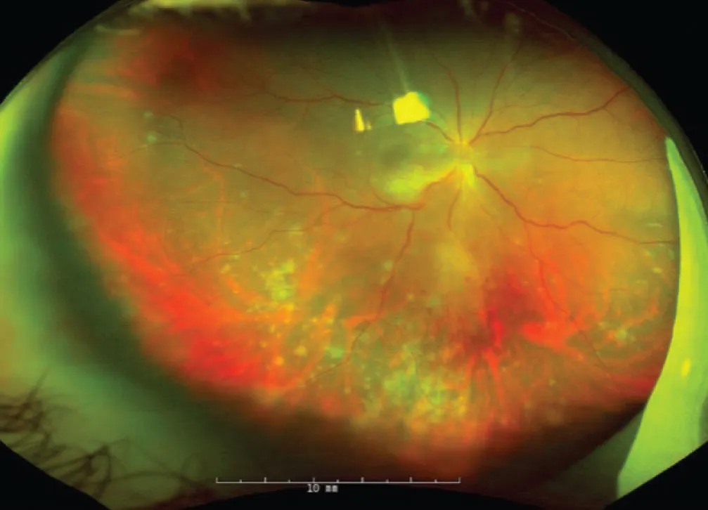
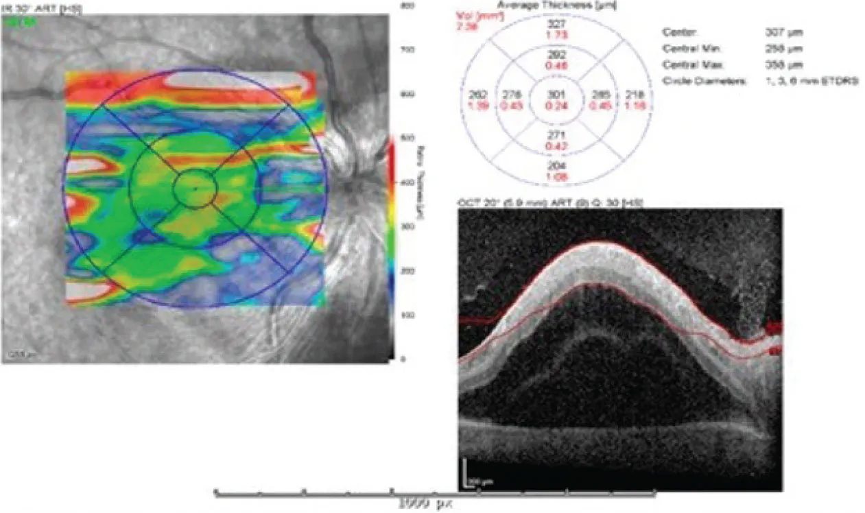
The patient received IV acyclovir 10 mg/kg every 8 hours with stabilization of retinitis and was transitioned to oral valacyclovir 1,000 mg every 8 hours in anticipation of discharge. Three days after initiation of valacyclovir, the patient developed an erythematous, maculopapular rash on left side of his face that spread to his arms, chest, and upper back. Biopsy revealed necrotic keratinocytes, consistent with a drug reaction. The patient’s rash gradually resolved after treatment with topical corticosteroids and cessation of valacyclovir. Due to concerns for recurrence, valganciclovir 450 mg twice daily was initiated. By day 60, the patient’s VA improved to 20/50 OD, and ophthalmic examination demonstrated resolution of the serous RD, peripheral granulated retinitis, and mild cystoid macular edema. The patient was discharged with close follow-up.
During outpatient care, the Infectious Diseases service transitioned him from valganciclovir to oral famciclovir 500 mg for lifelong suppression of HSV-2. Approximately 7 weeks after discharge, the patient woke up with redness and blurry vision in his right eye. At the outpatient clinic, his VA was 20/80 OD and his IOP was 55 mm Hg, and the retinitis had returned in the periphery (Figure 3). Given concern for treatment failure of famciclovir, repeat AC tap was performed for HSV-1, HSV-2, varicella zoster virus (VZV), and cytomegalovirus (CMV) DNA quantification and verified the presence of only HSV-2 by reverse transcription-PCR. He was subsequently readmitted for IV ganciclovir treatment.
During admission, antiviral therapy was escalated to combination IV and intravitreal foscarnet, given the potential disease progression on IV ganciclovir. As the retinitis resolved, patchy retinal breaks became apparent and were treated with laser retinopexy.
As the clinical examination stabilized during admission, the Infectious Disease service recommended re-trialing valacyclovir with weekly complete blood count and comprehensive metabolic panel monitoring. The patient tolerated valacyclovir and was discharged with resolving retinitis and a VA of 20/30 OD.
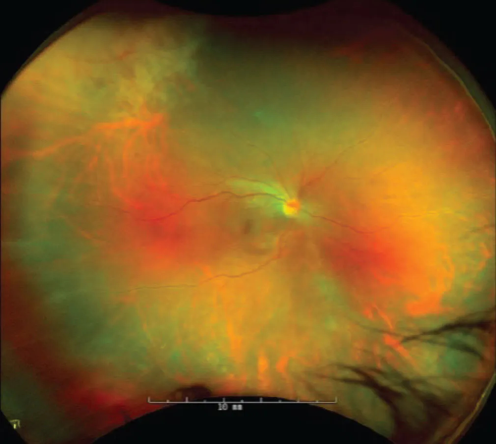
Figure 3. Fundus photography demonstrated the return of peripheral retinitis 4 months after initial presentation.
DISCUSSION
ARN is a type of infectious chorioretinitis, which is an inflammatory condition of the retina caused by an infectious pathogen.1 Common viral pathogens include HSV, VZV, CMV, and Epstein-Barr virus.1 Classically, VZV affects older individuals, while HSV affects younger patients.1 Certain genetic predispositions can increase a patient’s risk of developing ARN as well.2 ARN can affect both immunocompetent and immunocompromised patients.2
While the presentation of simultaneous BARN occurs in approximately one third of cases of ARN, delayed development of ARN in the initially unaffected eye is relatively low and can occur anywhere between 5 days to 30 years.3,4 However, reactivation more than 20 years later in the setting of immunocompetency is atypical. One study found the rate of ARN development in the initially unaffected eye while on maintenance antiviral therapy was 3.4%.5 Antiviral therapy significantly reduces the risk of bilateral involvement, with one study suggesting 87.1% of patients treated with acyclovir had fellow eyes that remained disease-free, compared with 30.4% of untreated patients.6 In our case, development of ARN in the right eye occurred approximately 30 years after ARN in the contralateral eye. This raises the question of the role of lifelong antiviral therapy in patients with a history of ARN while also considering that a single case cannot be used to generalize conclusions regarding guidelines for prophylaxis.
Managing ARN
Management of ARN can involve oral antivirals, IV antivirals, or combination therapy. One study demonstrated that visual acuity improvement was highest in patients who received combination therapy compared with oral or IV antivirals alone; however, the results were not statistically significant. Oral antiviral regimens include valacyclovir 1 g to 2 g three times daily until resolution of the retinitis, followed by a maintenance phase of valacyclovir tapered over 2 to 6 months or acyclovir 400 mg five times daily for HSV. Oral famciclovir can be given with a dose of 500 mg three times daily until resolution. Multimodal therapy can include the combination of either foscarnet and ganciclovir, oral famciclovir and intravitreal foscarnet, or intravenous acyclovir and intravitreal foscarnet with induction and maintenance phases.7
The risk of ARN recurrence while on antiviral maintenance therapy, while low, is possible and necessitates close follow-up. Resistance to antiviral drugs may contribute to the recurrence of HSV infections.8 There is no formal data on the recurrence rate of ARN on famciclovir maintenance therapy. The clinical course of our case suggests continued close monitoring of patients with ARN on famciclovir maintenance therapy is important, and adding alternative therapies—such as ganciclovir or foscarnet—in cases of recurrence may be necessary. Additionally, genotyping for HSV-2 resistant mutations was not performed, limiting the understanding of the disease recurrence, and should be considered in future cases of treatment failure.
Atypical Reaction to Valacyclovir
While effective, valacyclovir is not without side effects. The most common adverse reactions include headache, nausea, and abdominal pain, but it can rarely cause an erythematous and pruritic rash, particularly in those with HIV-1 infection.9 These reactions are generally mild but may require discontinuation of the medication in severe cases. In immunocompromised patients, valacyclovir can cause more severe dermatologic reactions, including symmetrical drug-related intertriginous and flexural exanthema, which typically affects areas such as the axillae and groin.10 The development of a rash nonresponsive to systemic steroids or antihistamines in an immunocompetent patient highlights the clinical challenge of managing this case.
A LONG HAUL FOR YOU AND THE PATIENT
ARN may present initially with isolated findings of optic nerve edema that may precede retinal findings. Patients with optic disc involvement should be monitored closely for additional findings that suggest an infectious etiology, particularly if they have a history of ocular viral infection. BARN can present in the setting of immunocompetency, and lifelong antiviral suppression may be warranted after an initial presentation of ARN. Side effects should be closely monitored in patients starting new antiviral therapy even in the absence of documented drug reactions.
1. Bergstrom R, Tripathy K. Acute retinal necrosis. In: StatPearls. StatPearls Publishing; 2024. Accessed September 3, 2026. www.ncbi.nlm.nih.gov/books/NBK470588
2. Coussa RG. Acute retinal necrosis. EyeWiki. February 1, 2026. Accessed September 3, 2026. tinyurl.com/mvrwue93
3. Nguyen NM, Conrady CD. A better understanding of the clinical and pathological changes in viral retinitis: steps to improve visual outcomes. Microorganisms. 2024;12(12):2513. doi.org/10.3390/microorganisms12122513
4. Mitkova-Hristova V, Stoyanova NS. Acute retinal necrosis: pathophysiological aspects, diagnosis, and treatment. Folia Med (Plovdiv). 2022;64(6):871-877. doi.org/10.3897/folmed.64.e68036
5. Tibbetts MD, Shah CP, Young LH, Duker JS, Maguire JI, Morley MG. Treatment of acute retinal necrosis. Ophthalmology. 2010;117(4):818-824. doi.org/10.1016/j.ophtha.2009.09.001
6. Palay DA, Sternberg P, Davis J, et al. Decrease in the risk of bilateral acute retinal necrosis by acyclovir therapy. Am J Ophthalmol. 1991;112(3):250-255. doi.org/10.1016/s0002-9394(14)76725-x
7. Putera I, Ridwan AS, Dewi M, et al. Antiviral treatment for acute retinal necrosis: A systematic review and meta-analysis. Surv Ophthalmol. 2024;69(1):67-84. doi.org/10.1016/j.survophthal.2023.09.004
8. National Library of Medicine. VALTREX- valacyclovir hydrochloride tablet, film coated. Updated July 16, 2026. Accessed September 3, 2026. tinyurl.com/s9z2bs7p
9. Daito J, Hanada K, Katoh N, et al. Symmetrical drug-related intertriginous and flexural exanthema caused by valacyclovir. Dermatol Basel Switz. 2009;218(1):60-62. doi.org/10.1159/000167829
10. Sharma A, Roy S, Sharma R, Kumar A. Association of antiviral drugs and their possible mechanisms with DRESS syndrome using data mining algorithms. J Med Virol. 2023;95(3):e28671. doi.org/10.1002/jmv.28671
11. Ng CC, McDonald HR, Johnson RN, Cunningham ET. Optic disc edema with peripapillary serous retinal detachment as the presenting sign of necrotizing herpetic retinitis. Am J Ophthalmol Case Rep. 2022;25:101423. doi.org/10.1016/j.ajoc.2022.101423


