


KEY TAKEAWAYS
- Although many low- and middle-income countries have expanded neonatal intensive care unit capabilities and improved preterm infant survival rates, health care infrastructure and resources to screen premature infants at risk of retinopathy of prematurity (ROP) have lagged.
- A pilot telemedicine ROP (teleROP) screening program was launched in 2024 as a partnership between experts at Stanford University and the Komfo Anokye Teaching Hospital and was later expanded across Ghana and Ethiopia.
- The program has noted region-associated pathological distinctions in ROP; while only 7.5% of neonates at Standford University had treatment-warranted ROP driven by zone I stage 3 without plus disease, 40% of treated eyes in Ghana had treatment indications based on zone-driven disease.
Retinopathy of prematurity (ROP) is a preventable condition that remains the leading cause of childhood blindness.1,2 In the 21st century, the third ROP epidemic has disproportionately affected low- and middle-income countries (LMICs), including many in sub-Saharan Africa. Many LMICs have expanded neonatal intensive care unit (NICU) capabilities and improved preterm infant survival rates, but health care infrastructure and resources to screen premature infants at risk of blindness from ROP have lagged.3 Particularly in NICUs in rural areas, premature infants are still exposed to unfractionated oxygen at high rates due to limited numbers of oxygen blenders and continuous positive airway pressure machines. Access to ophthalmologists who can screen and treat ROP is also limited; in Ghana and Ethiopia, ophthalmologists are already in short supply, much less specialists in pediatric ophthalmology or retina.4,5
TELEMEDICINE PROGRAM TO ENHANCE ROP SCREENING
To bridge gaps in ROP screening, telemedicine-based ROP (teleROP) screening has been adopted and validated throughout LMICs and high-income settings alike, providing improved access within a country due to the concentration of specialists in major urban areas.6 In the United States, the Stanford University Network for Diagnosis of Retinopathy of Prematurity (SUNDROP) initiative began in 2005 and has been validated to have high sensitivity and specificity for treatment-warranted ROP (TW-ROP).7 Since 2007, the Karnataka Internet Assisted Diagnosis for Retinopathy of Prematurity model has successfully been screening infants throughout rural and semi-urban India with nonphysician imagers/graders who travel an average of 7,000 km each month.8
A pilot teleROP screening program was launched in 2024 as a partnership between quaternary teleROP experts at Stanford University and the Komfo Anokye Teaching Hospital. The model was then refined and scaled to three other metropolitan areas: Accra and Tamale in Ghana and Addis Ababa in Ethiopia. In the first 22 months of the program, more than 3,000 infants have been screened over nearly 13,000 visits, generating more than 185,000 images and treating 92 infants.
Such a program has been accomplished through the diligence of a team that includes eight full-time, in-country ROP coordinators and imagers, two country-wide managers, a network of local physicians (neonatologists, vitreoretinal surgeons, and pediatric ophthalmologists), and a quaternary teleROP expert at Stanford University.
After referral of infants meeting local ROP screening criteria (birth weight ≤ 2,000 g or gestational age ≤ 32 weeks in Ghana; birth weight ≤ 2,500 g or gestational age ≤ 34 weeks in Ethiopia; or medical course deemed high-risk by a local neonatologist), widefield fundus images are captured with a 120° or 130° camera. The images are graded by a local ophthalmologist within 24 hours and concurrently by a quaternary teleROP expert at Stanford University to determine appropriate follow-up. Infants deemed to have TW-ROP undergo treatment managed by the local ROP coordinators, typically initially with intravitreal bevacizumab (Avastin, Genentech/Roche), until laser to the peripheral avascular retina can be completed at an older age.
DISTINCT PATHOLOGIES ACROSS REGIONS
The pathology of ROP at the sites in Ghana and Ethiopia varied slightly from that which has been noted at Stanford University and in high-income countries generally, likely due to variations in neonatal practices in sub-Saharan African NICUs, although an underlying genetic predisposition to different phenotypes of ROP cannot be excluded.
Case Example
An infant was born at 26 5/7 weeks estimated gestational age with a birth weight of 1,000 g at an outside private NICU where inpatient ROP screening was not routinely performed because of the cost and logistical burden of transporting the screening team and widefield retinal camera. Although the infant ideally would have been screened at 30 weeks’ postmenstrual age based on Ghana screening criteria, she presented through the outpatient referral network at 37 4/7 weeks’ postmenstrual age with TW-ROP, including zone I plus disease in each eye (Figure A). She was treated with bilateral intravitreal bevacizumab at 37 5/7 weeks’ postmenstrual age, with regression at 38 4/7 weeks (Figure B) and further regression at 40 4/7 weeks (Figure C). She later developed reactivation at 43 4/7 weeks’ postmenstrual age (Figure D), underwent bilateral laser photocoagulation at 44 5/7 weeks, and was quiescent at 57 4/7 weeks (Figure E).
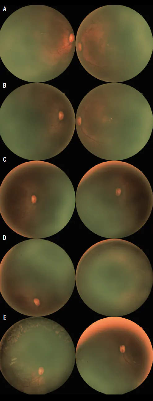
Figure. An infant presented at 37 4/7 weeks’ postmenstrual age with zone I plus disease in each eye (A). Bilateral intravitreal bevacizumab led to regression at 38 4/7 weeks (B) and 40 4/7 weeks (C). Reactivation was noted at 43 4/7 weeks (D), prompting bilateral laser photocoagulation. The eyes were quiescent at 57 4/7 weeks (E).
This case demonstrates that even delayed outpatient referral-based screening can identify and treat severe ROP in settings where universal inpatient and outpatient screening is not yet feasible.
The Need for Region-Specific Recommendations
Of note, this patient would not have met US ROP screening criteria by either her gestational age (≤ 30 weeks) or birth weight (≤ 1,500 g). These differences in ROP patterns across different international settings highlight the need for investigation of region-specific epidemiology and adoption of recommendations determined by local ROP experts.
As a corollary, a significant difference this program has noticed between the infant eyes treated in Ghana versus those treated in SUNDROP (ie, in the United States) is the absence of plus disease as a driver of TW-ROP. While only 7.5% of neonates in SUNDROP had TW-ROP driven by zone I stage 3 without plus disease, 40% of treated eyes in Ghana had treatment indications based on zone-driven disease.9 As has also been noticed in other LMICs,10,11 treated infants in Ghana have also been significantly older and heavier at birth.9 Aggressive ROP (AROP), which classically presents with plus disease, rapid neovascular proliferation, and lack of typical stage progression, has been observed in different phenotypes, which we have termed typical minus plus-AROP and placoid minus plus-AROP.
Another key difference is the outpatient setting in which much of neonatal care takes place. Unlike in many high-income countries, Ghana and Ethiopia have a care delivery model that relies heavily on follow-up by families from the outpatient setting. Due to resource capacity, many infants are discharged from the NICU shortly after they are weaned off oxygen, often at 34 to 35 weeks, the period during which they are at highest risk for active ROP. Hence, caregiver education, robust referral systems, and careful tracking for compliance by ROP coordinators is crucial.
ROP Grading Models
To simultaneously build local capacity for ROP screening and treatment, this program employs a dual-layer telemedicine grading model. Local ophthalmologists do the initial grading, and a teleROP expert at Stanford University contemporaneously validates the results within 24 hours. This model allows both the timely clinical decision making and parental counseling needed for patient safety and care, as well as increased autonomy of local graders over time.
AI models show promise to eventually improve screening and triaging efficiency,12 but thorough validation will be required, especially given the regional variations in pathology.
As the program continues to reach infants at risk of blindness from ROP, its success hinges on partnerships with local physicians and an emphasis on the need for continued follow-up with parents. Treatments are readily available and often used in the same settings for adult diseases by retina specialists throughout the world. Different models may be required to adapt to unique settings; currently, our program in Ghana has a hub-and-spoke referral network, while the one in Ethiopia employs a mobile screening team.
From the perspective of cost, identifying and addressing TW-ROP at a total program cost of $450 per treated infant (~$50 for bilateral intravitreal bevacizumab) is negligible compared with the societal burden of childhood blindness.
PREVENT ROP WORLDWIDE
Through sufficient screening, improved awareness of pediatricians and neonatologists in sub-Saharan Africa, and increasing the capacity of local ophthalmologists, ROP, a preventable disease, does not need to be the leading cause of infant blindness.
1. Blencowe H, Lawn JE, Vazquez T, Fielder A, Gilbert C. Preterm-associated visual impairment and estimates of retinopathy of prematurity at regional and global levels for 2010. Pediatr Res. 2013;74 Suppl 1(Suppl 1):35-49. doi.org/10.1038/pr.2013.205
2. Lim HW, Pershing S, Moshfeghi DM, Heo H, Haque ME, Lambert SR; IRIS Registry Analytic Center Consortium. Causes of childhood blindness in the United States using the IRIS Registry (Intelligent Research in Sight). Ophthalmology. 2023;130(9):907-913. doi.org/10.1016/j.ophtha.2023.04.004
3. Gilbert C, Malik ANJ, Nahar N, et al. Epidemiology of ROP update - Africa is the new frontier. Semin Perinatol. 2019;43(6):317-322. doi.org/10.1053/j.semperi.2019.05.002
4. Mehari Z, Nariani A. Assessment of human resource for eye health services in Southern Ethiopia. Hum Resour Health. 2025;23(1):52. doi.org/10.1186/s12960-025-01007-1
5. Dean WH, Buchan JC, Gichuhi S, Faal H, Mpyet C, Resnikoff S, et al. Ophthalmology training in sub-Saharan Africa: a scoping review. Eye (Lond). 2021;35(4):1066-1083. doi.org/10.1038/s41433-020-01335-7
6. Quinn GE, Vinekar A. The role of retinal photography and telemedicine in ROP screening. Semin Perinatol. 2019;43(6):367-374. doi.org/10.1053/j.semperi.2019.05.010
7. Wang SK, Callaway NF, Wallenstein MB, Henderson MT, Leng T, Moshfeghi DM. SUNDROP: six years of screening for retinopathy of prematurity with telemedicine. Can J Ophthalmol. 2015;50(2):101-106. doi.org/10.1016/j.jcjo.2014.11.005
8. Vinekar A, Mangalesh S, Jayadev C, Gilbert C, Dogra M, Shetty B. Impact of expansion of telemedicine screening for retinopathy of prematurity in India. Indian J Ophthalmol. 2017;65(5):390-395. doi.org/10.4103/ijo.IJO_211_17
9. Shah SV, Brant AR, Ahmed A, et al. Regional variations in type 1 retinopathy of prematurity: GHANAROP and SUNDROP. [published online ahead of print April 6, 2026.] Br J Ophthalmol. doi.org/10.1136/bjo-2025-328512
10. Sanghi G, Dogra MR, Das P, Vinekar A, Gupta A, Dutta S. Aggressive posterior retinopathy of prematurity in Asian Indian babies: spectrum of disease and outcome after laser treatment. Retina. 2009;29(9):1335-1339. doi.org/10.1097/IAE.0b013e3181a68f3a
11. Shah PK, Narendran V, Kalpana N. Aggressive posterior retinopathy of prematurity in large preterm babies in South India. Arch Dis Child Fetal Neonatal Ed. 2012;97(5):F371-375. doi.org/10.1136/fetalneonatal-2011-301121
12. Placide J, Coyner AS, Ostmo SR, et al. Precision risk model using quantitative assessment of vascular severity in telemedicine-based screening [published online ahead of print April 2, 2026]. JAMA Ophthalmol. doi.org/10.1001/jamaophthalmol.2026.0510


