




KEY TAKEAWAYS
- Thorough vitreous base shaving is essential to prevent the formation of postoperative proliferative vitreoretinopathy following retinal detachment repair.
- In phakic eyes, vitreous base shaving and peripheral endolaser are often challenging due to difficulties in assessing the depth of the surgical instruments relative to the crystalline lens, poor visibility of the peripheral vitreous, and complex visualization under air.
- The authors describe an approach termed the bubble technique that may provide useful visual cues to assess the depth of the instrument relative to the crystalline lens, thus avoiding iatrogenic complications while allowing good peripheral visualization under air.
Vitreous base shaving is essential for a successful surgical outcome in retinal detachment (RD) repair.1 However, this maneuver can be challenging in phakic eyes. Primary hurdles include difficulty assessing the instrument depth relative to the crystalline lens, limited visibility of the extreme periphery, and navigating complex visualization under air. To address these specific issues, we propose a maneuver we have termed the bubble technique.
THE IMPORTANCE OF VITREOUS BASE SHAVING
Residual vitreous base can act as a scaffold for proliferative vitreoretinopathy (PVR) formation after RD repair, and subsequent contraction of the remnant vitreous can lead to formation of new breaks.1,2 Therefore, thorough base shaving is essential. The goal is to remove as much vitreous as possible without causing iatrogenic breaks.
To achieve this, surgeons can employ scleral indentation by the surgical assistant or use chandelier illumination to allow indentation by the surgeon. A wide-angle viewing system enables visualization of peripheral retina without the need for indentation. In addition, employing a high cut rate and low vacuum facilitates controlled removal of vitreous base. The advantages of vitrectomy under air are well known.3
THE BUBBLE TECHNIQUE: STEP-BY-STEP
To shave the vitreous base in phakic eyes while maintaining good visualization, surgeons can proceed with the following steps:
- Perform standard pars plana vitrectomy and induce a posterior vitreous detachment, if not already present.
- Complete a fluid-air exchange with subretinal fluid drainage with or without the assistance of perfluorocarbon liquid.
- Restart the fluid infusion to achieve an intentional, incomplete removal of air. The residual air bubble should be single and large enough to allow clear visualization of the periphery (Figure).
- Upon scleral indentation, the air bubble dynamically occupies the area between the indented retina and the posterior lens capsule. This configuration places the air bubble above the fluid, which facilitates excellent peripheral visualization while maintaining critical depth perception for safe use of instruments. Note: This adjunctive step is intended for final base shaving only after initial peripheral debulking has been completed under fluid. Although the air tamponade compresses the residual vitreous closer to the retina, the resulting optical interface clearly demarcates the tissue boundaries, facilitating safe and precise removal. Importantly, this technique does not alter the physical constraints of the phakic lens and does not safely permit "crossing" the eye with instruments. To address this, a skilled assistant must indent the ora serrata with a posterior "rolling" motion, rather than direct inward pressure; this method safely displaces the vitreous base posteriorly and inward toward the central cavity, making it safe to approach. Finally, because the vitrectomy cutter inevitably aspirates air despite low vacuum settings, the surgeon must actively monitor and occasionally supplement the bubble volume to maintain optimal visualization.
- Apply endolaser in difficult peripheral areas concurrently during the base shaving process in the presence of the air bubble. Upon successful completion of vitreous base shaving, proceed with routine endolaser and final endotamponade.
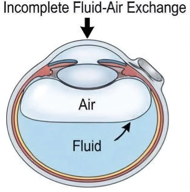
Figure. Illustration of the bubble technique: An incomplete fluid-air exchange maintains a buoyant anterior air bubble above the fluid, enabling peripheral visualization, while the distinct fluid-air meniscus provides crucial depth perception to prevent lens touch. Note: The author used ibispaintx to create the image and Gemini 3.1 Pro (Google) to refine it.
UNDERSTANDING THE OPTICS
The success of this technique relies heavily on fluid dynamics and optical physics. Because the specific gravity of air is less than that of balanced salt solution, air exhibits high buoyancy. In a supine position, this buoyancy causes the air to float in the anterior vitreous cavity in the presence of fluid. Dynamic scleral indentation displaces the peripheral retina inward bringing the target area into the accessible surgical field within the air medium. As the light rays reflect off the retina and exits the eye, they travel from the air medium (refractive index = 1.00) into the crystalline lens (refractive index = 1.40). Because of this large difference in refractive index at the convex posterior lens capsule, the posterior capsule acts as a strong converging optical surface, causing the extreme peripheral light rays to bend inward and allowing them to pass through the pupillary aperture. Thus, the bubble technique can overcome standard visual hindrances, such as the iris. Furthermore, the fluid-air interface acts as an optical reference plane, providing visual cues to gauge the exact location of the cutter or laser probe relative to the lens.
USE PHYSICS TO YOUR ADVANTAGE
Employing the bubble technique facilitates vitreous base shaving while effectively avoiding complications, such as lens touch, and affording the surgeon the distinct advantage of excellent peripheral visualization under air. However, the surgeon must be aware of the limitations associated with this technique, such as the dynamic fluidics altering the bubble volume and the proximity of the vitreous to the retina under air.
1. Chaturvedi V, Basham RP, Rezaei KA. Scleral depressed vitreous shaving, 360 laser, and perfluoropropane (C3F8) for retinal detachment. Indian J Ophthalmol. 2014;62(7):804-808. doi.org/10.4103/0301-4738.138621
2. Nishitsuka K, Nakamura M, Nishi K, Namba H, Kaneko Y, Yamashita H. Surgical outcomes of rhegmatogenous retinal detachment with different peripheral vitreous-shaving procedures. Clin Ophthalmol. 2021;15:2197-2202. doi.org/10.2147/OPTH.S310789
3. Voleti VB, Gee CJ, Devin F, Hubschman JP. Vitrectomy under air. Retina. 2012;32(9):1981-1982. doi.org/10.1097/IAE.0b013e31826711a9


