





KEY TAKEAWAYS
- Despite substantial achievements in expanding the treatment of uveal melanoma beyond enucleation, it is important to recognize that preserving the eye does not necessarily mean preserving vision.
- In a cohort analysis, the authors found that vision loss after plaque radiotherapy often began early: 30% of eyes lost at least 15 letters by 1 year, 48% by 2 years, and 60% by 3 years.
- Regarding effects of radiation dose, keeping the optic disc dose at or below 4,000 cGy and the foveolar dose below 2,000 cGy appeared to offer the best chance of visual preservation.
The management of posterior uveal melanoma has undergone a remarkable journey. The first battle was directed toward saving the patient’s life with detection of uveal melanoma when the tumor is smaller and genetic information demonstrates fewer high-risk mutations.1,2
In the past, enucleation was the only treatment option. Today, plaque radiotherapy and proton beam radiotherapy are widely used, while gamma knife and CyberKnife (Accuray) radiotherapy are available in select centers. These globe-conserving therapies allow preservation of 85% of eyes with uveal melanoma and achieve excellent local tumor control, with recurrence rates of only 2% to 5%.3 Despite these achievements, preserving the eye does not necessarily mean preserving vision.
Even with successful local tumor control, radiation maculopathy, radiation papillopathy, and other radiation-related complications remain principal causes of long-term visual impairment. Months or even years after treatment, radiation can progressively damage the macula and optic nerve, leading to irreversible vision loss (Figure). The current battle is therefore not simply to control the tumor, prevent metastasis, and retain the globe, but also to preserve functional vision whenever possible.
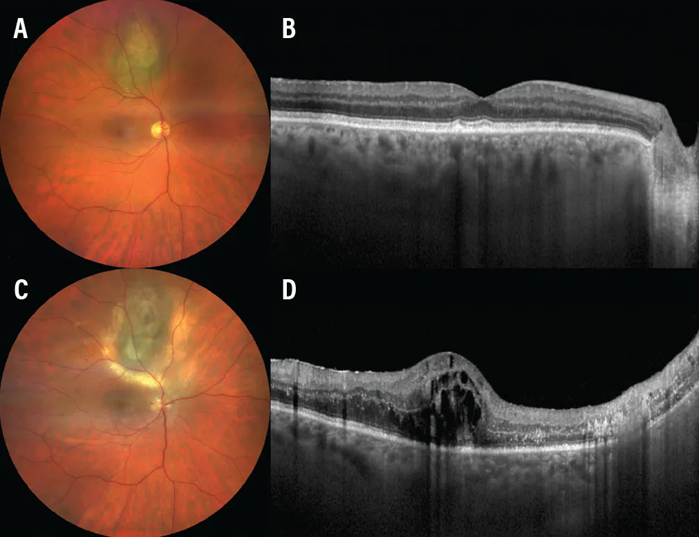
Figure. Multimodal imaging of a superotemporal choroidal melanoma in the right eye of a patient before and after plaque radiotherapy. Baseline color fundus photography demonstrates a superotemporal choroidal melanoma in the right eye (A). Corresponding OCT at baseline shows a preserved foveal contour without cystoid macular edema or subretinal fluid (B). Color fundus photography obtained 2 years after plaque radiotherapy shows the regressed superior paramacular tumor with surrounding retinal exudation and retinal pigment epithelium atrophy, additional cotton-wool spots at the optic disc, optic disc hemorrhage, and edema, consistent with radiation papillopathy. Also note the presence of numerous scattered dot hemorrhages, predominantly involving the superotemporal retina, consistent with radiation retinopathy (C). Corresponding OCT obtained at the same follow-up visit demonstrates severe cystoid macular edema and loss of the normal foveal contour, consistent with radiation maculopathy (D).
VISION DECLINE AFTER PLAQUE RADIOTHERAPY
We performed an analysis of 4,000 patients with plaque-irradiated uveal melanoma who experienced a loss of at least 15 letters of vision, roughly 3 lines on the visual acuity chart.4 All patients were managed in the pre-anti-VEGF era, so there were no injections or treatments for vision decline. We found that vision loss often began early: 30% of eyes lost at least 15 letters within 1 year, 48% by 2 years, and 60% by 3 years. This increased to 73% at 5 years and 82% at 10 years. Overall, vision loss occurred at a mean of approximately 25 months, suggesting the first few of years after treatment are a particularly important period for close monitoring.4
Tumor size also mattered. Larger tumors were associated with earlier and more frequent vision loss of at least 15 letters, occurring in 56% of small tumors (0 to 3 mm in thickness), 62% of medium tumors (3.1 mm to 8 mm), and 70% of large tumors (> 8 mm) over a mean follow-up period of 69 months. For small tumors, vision loss occurred at a mean of 33 months, for medium tumors at a mean of 25 months, and for large tumors at a mean of 13 months. Put simply, visual decline after plaque radiotherapy is common, accumulates over time, and occurs more rapidly in eyes with larger tumors.4
PATIENTS AT RISK FOR VISION DECLINE
Our second analysis of this cohort asked a practical question: Can we predict which patients are most likely to experience meaningful vision loss? On multivariate analysis, the most important risk factors for vision loss were greater tumor thickness and closer proximity to the foveola.4 Each additional millimeter of tumor thickness increased the risk of vision loss by approximately 14%, and each millimeter farther from the foveola reduced the risk by approximately 7%.4 Having diabetes also carried an independent increased risk for vision loss. In addition, treatment-related factors were significant, particularly greater radiation dose to the foveola and lens.
These findings divide risk into three categories: presence of comorbidities, including diabetes; characteristics of the tumor, particularly thickness and location; and the specific treatment, ie, radiation exposure to visually important structures. Tumor thickness remained particularly important for medium and large tumors, whereas foveolar proximity was most relevant for small and medium tumors. This gives clinicians a straightforward way to counsel patients: Thicker tumors, tumors close to central vision, diabetes, and greater radiation exposure all lead to a greater risk of post-treatment visual decline.4
Of note, many of these determinants are established well before treatment begins, including tumor thickness, location of the tumor relative to the foveola and optic disc, and patient characteristics. Although these factors cannot be changed, radiation exposure remains at least partially within our control.
RADIATION DOSE MATTERS
The third analysis we performed for this cohort focused on radiation dose to the optic disc and foveola.5 There was no absolute dose at which vision was suddenly lost; rather, the study demonstrated a clear dose response relationship. When the optic disc received no more than 4,000 cGy, increasing the foveolar dose from ≤ 2,000 cGy to 2,001 cGy to 4,000 cGy was associated with a 71% higher risk of losing at least 15 letters. Increasing the foveolar dose further to 4,001 cGy to 6,000 cGy produced an additional increase in risk. When the optic disc dose exceeded 4,000 cGy, visual outcomes were generally poor, regardless of the foveolar dose. Thus, keeping the optic disc dose at or below 4,000 cGy and the foveolar dose below 2,000 cGy appears to offer the best chance of visual preservation. Careful treatment planning, including precise plaque and seed positioning, may help reduce radiation exposure to critical structures whenever tumor location allows.5
A novel strategy being explored is to reduce tumor size before plaque radiotherapy. Neoadjuvant therapy could permit lower radiation exposure to the foveola and optic disc.6,7 Darovasertib, an oral protein kinase C inhibitor, has shown early evidence for tumor reduction before definitive treatment of primary uveal melanoma.7 Tebentafusp improves overall survival in HLA-A*02:01 positive metastatic uveal melanoma, but it has yet to show efficacy as a neoadjuvant tumor-shrinking therapy.8
Although still investigational, neoadjuvant therapy to reduce tumor size prior to radiation may represent a new paradigm that could expand opportunities not only for globe preservation, but also for vision preservation.
WHAT COMES NEXT?
Our three studies sought to define how often vision is lost, which patients are most vulnerable, and how radiation dose affects risk.4,5 They also provide a large, historical pre-anti-VEGF cohort for comparison with newer strategies. Our group is now studying prophylactic intravitreal anti-VEGF injections after plaque radiotherapy to determine whether scheduled treatment can reduce radiation-related retinal damage and vision loss.
We have learned how to save the patient’s life with early detection of tumor,1,2 and we have learned how to save their eyes using plaque radiotherapy.3 Now, we must learn how to save their sight.
Acknowledgements: Support provided in part by the Jerry A. Shields, MD, Eye Cancer Fund, Philadelphia, PA (CLS), the Eye Tumor Research Foundation, Philadelphia, PA (CLS) and the Melanoma Research Foundation (ABG). The funders had no role in the design and conduct of the study, in the collection, analysis, and interpretation of the data, and in the preparation, review or approval of the manuscript. Carol L. Shields, MD, has had full access to all the data in the study and takes responsibility for the integrity of the data.
AI disclosure: ChatGPT (GPT-5.6 Sol, OpenAI), was used to assist with language editing and manuscript organization. All content was reviewed, verified, and revised by the authors, who assume full responsibility for the accuracy and integrity of the manuscript. The AI tool was not used for data analysis, interpretation, or drawing scientific conclusions.
1. Shields CL, Furuta M, Thangappan A, et al. Metastasis of uveal melanoma millimeter by millimeter in 8033 consecutive eyes. Arch Ophthalmol. 2009;127(8):989-998. doi.org/10.1001/archophthalmol.2009.208
2. Shields CL, Say EAT, Hasanreisoglu M, et al. Cytogenetic abnormalities in uveal melanoma based on tumor features and size in 1059 patients. Ophthalmology. 2017;124(5):609-618. doi.org/10.1016/j.ophtha.2016.12.026
3. Shields CL, Cater J, Shields JA, et al. Combined plaque radiotherapy and transpupillary thermotherapy for choroidal melanoma: tumor control and treatment complications in 270 consecutive patients. Arch Ophthalmol. 2002;120(7):933-940. doi.org/10.1001/archopht.120.7.933
4. Shields CL, Medina RJ, Sener H, et al. Plaque radiotherapy for posterior uveal melanoma in 4000 eyes: analysis of ≥15-letter visual acuity loss in the pre-anti-VEGF era. Retina. 2026;46(8):1363-1370. doi.org/10.1097/IAE.0000000000004849
5. Shields CL, Medina RJ, Sener H, et al. Plaque radiotherapy for posterior uveal melanoma in 4000 eyes: Impact of radiation dose to the optic disc and foveola on 15 letter visual acuity loss in the pre anti-VEGF era. Retina. In press.
6. Shields CL. Enucleation prevention and vision preservation in primary uveal melanoma (UM): Preliminary results from a phase 2 study of neoadjuvant Darovasertib. Poster presented at: 44th Annual Meeting of the American Society of Retina Specialists; July 15-18, 2026; Montreal, Quebec, Canada.
7. Hiong A, O’Day R, Fog LS, et al. Globe salvage and vision preservation by neoadjuvant Darovasertib and Crizotinib in uveal melanoma. Ophthalmol Retina. 2024;8(4):325-330. doi.org/10.1016/j.oret.2023.10.009
8. Nathan P, Hassel JC, Rutkowski P, et al. Overall survival benefit with Tebentafusp in metastatic uveal melanoma. N Engl J Med. 2021;385(13):1196-1206. doi.org/10.1056/NEJMoa2103485


