



Geographic atrophy (GA) is increasingly recognized as a progressive disease that warrants intervention before central vision is irreversibly compromised. However, defining “early” GA remains a clinical challenge. In my clinic, GA lesions that are smaller than 1 disc diameter of total atrophy are considered early disease. However, diagnosing by lesion size alone is insufficient. Lesion location, progression, and the presence of multifocal lesions also inform decision-making, as real-world treatment decisions often require a more nuanced assessment of disease trajectory and functional risk.
 Defining Early GA: More Than Lesion Size
Although literature supporting treatment at very early stages remains limited, the inclusion criteria used in the pivotal OAKS/DERBY and GATHER 1 and 2 clinical trial programs provide a framework for identifying patients that will likely benefit from treatment.1-3 In these trials, enrollment included patients with at least 1 disc area of GA, or approximately 2.5 mm² with a maximum lesion size of 17.5 mm², while excluding those patients with contiguous peripapillary atrophy. Multifocal lesions that added up to 2.5 mm² were also allowed if one GA lesion was at least 1.25 mm² and the other lesions totaled 2.5 mm². Smaller lesions may present challenges for treatment decisions; larger lesions or those that have peripapillary atrophy, not involving the fovea, do not present as much of a therapeutic challenge.
 Recognizing Early Disease Through Imaging
In addition to the clinical examination, I collect OCT images, which serve as a practical baseline tool for identifying early atrophic changes. When reviewing these images, I look for complete or incomplete retinal pigment epithelium and outer retinal atrophy, hyperreflective foci, and ellipsoid zone disruption. Often on the SD-OCT, there are hypertransmission defects that alert the clinician to problematic areas. Fundus autofluorescence typically defines GA more clearly than color photography and is the basis of measurement for the pivotal GA trials.1-3(Figure)
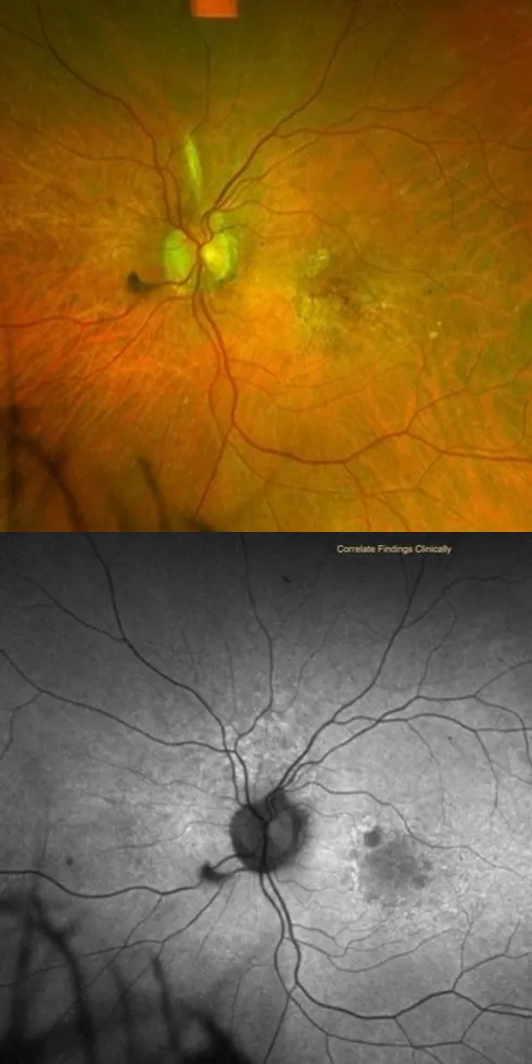
Figure. Fundus autofluorescence (FAF) imaging (bottom) often depicts areas of atrophy that are difficult to discern on color fundus photography (top). In these images, note how the borders of the emerging lesion are unclear on fundus photography, whereas they can be more clearly appreciated on FAF.
 Deciding When to Treat: Progression, Risk, and Functional Impact
Treatment discussions may be reasonable when small lesions threaten central vision. Beyond lesion size, I also consider several factors before making treatment decisions:
- Evidence of progression over time
- Foveal proximity or threat
- Fellow-eye vision loss from GA
- Patient-reported visual symptoms
- Functional limitations during reading or daily activities
In my experience, a strong patient candidate for receiving treatment for early disease is someone with vision loss in the fellow eye from end-stage wet AMD or foveal GA, with early atrophy developing in the functional eye. Patients who are experiencing noticeable visual changes are also strong candidates for treatment. While AREDS supplementation remains part of management discussions, I also discuss the possible benefits of photobiomodulation (PBM), which may be considered in select patients with intermediate AMD or even early atrophic changes.4 One study showed that GA incidence decreased from 24.0% in sham eyes to 6.8% in PBM- treated eyes (P = .007).
Unlike wet AMD, GA generally allows time for observation and shared decision-making between doctor and patient. To this end, repeat imaging at 3- to 6-month intervals may aid decision-making for borderline cases.
 Preserving Vision Over Time
There has been some resistance in the retina community when it comes to treating GA, with some doubting how much benefit we are truly providing. For patients who are anxious about treatment, I reference one recent study with long-term data demonstrating that, at 5 years, treatment preserved approximately a year and a half's worth of retinal tissue.5 Ultimately, long-term treatment for GA should be viewed similarly to the management of other chronic diseases, with the goal of preserving retinal tissue and visual function over time rather than restoring lost vision.
1. Heier JS, Lad EM, Holz FG, et al; OAKS and DERBY Study Investigators. Pegcetacoplan for the treatment of geographic atrophy secondary to age-related macular degeneration (OAKS and DERBY): two multicentre, randomised, double-masked, sham-controlled, phase 3 trials. Lancet. 2023;402(10414):1434-1448. doi: 10.1016/S0140-6736(23)01520-9.
2. Patel SS, Lally DR, Hsu J, et al. Avacincaptad pegol for geographic atrophy secondary to age-related macular degeneration: 18-month findings from the GATHER1 trial. Eye (Lond). 2023;37(18):3551-3557. doi: 10.1038/s41433-023-02548-2.
3. Khanani AM, Patel SS, Staurenghi G, et al. Efficacy and safety of avacincaptad pegol in patients with geographic atrophy (GATHER2): 12-month results from a randomized, double-masked, phase 3 trial. Lancet. 2023;402(10410):1449-1458.doi: 10.1016/S0140-6736(23)01583-0.
4. Boyer D, Hu A, Warrow D, et al. LIGHTSITE III: 13-month efficacy and safety evaluation of multiwavelength photobiomodulation in nonexudative (dry) age-related macular degeneration using the Lumithera Valeda Light delivery system. Retina. 2024;44(3):487-497. doi: 10.1097/IAE.0000000000003980.
5. Dhoot D, Garg S, Lally D, et al. Early treatment yields better outcomes: 5 years of pegcetcoplan treatment for geographic atrophy secondary to AMD. The Macula Society Annual Meeting. Feb 25-28, 2026. Coronado, CA.


