





KEY TAKEAWAYS
- Geometric perfusion deficit (GPD) on OCT angiography (OCTA) defines nonperfused retinal tissue based on the distance threshold from the nearest perfused capillary.
- In a prospective study, GPD in the deep capillary plexus was the only OCTA metric to detect significant microvascular changes in eyes with referable diabetic retinopathy (DR) at 6 months, ahead of other metrics that became significant only at 12 months.
- Higher baseline deep capillary plexus GPD predicted earlier onset of DR complications.
- OCTA-derived GPD-deep capillary plexus may complement conventional DR grading.
The current staging framework for diabetic retinopathy (DR) triages patients to broad severity categories; however, within a given severity category, the framework does not necessarily identify eyes with greater risk of progression to vision-threatening complications.1,2
Retinal ischemia is central to DR pathophysiology, with ischemic burden being an important key in defining risk of progression in diabetic eyes.3
Classically, ultra-widefield fluorescein angiography (UWF-FA) is used to document the extent of retinal nonperfusion, but this dye-based tool does not lend itself to routine or frequent imaging.1,4
In contrast, OCT angiography (OCTA) enables noninvasive, depth-resolved quantification of retinal capillary perfusion at the microvascular level, without impedance related to dye or leakage artifacts. Although OCTA images may capture only a portion of the total retinal area accessible to UWF-FA, the technology is rapidly evolving in speed and precision, allowing clinicians to image wider areas of the retina.
Furthermore, emerging evidence suggests that OCTA biomarkers may complement conventional DR staging in terms of the retinal ischemic burden.5
QUANTIFYING ISCHEMIA WITH OCTA
OCTA has the advantage of enabling layer-specific evaluation of retinal capillary perfusion. Its depth-resolved capability is particularly relevant, as diabetic capillary damage may not occur uniformly across retinal layers.6 Quantitative metrics of OCTA-based capillary perfusion have emerged as a useful approach to characterize diabetic retinal ischemia.7,8
One such metric is geometric perfusion deficit (GPD), which defines the proportion of ischemic retinal tissue farther than 30 µm from the nearest perfused capillary (Figure 1), or the theoretical oxygen diffusion limit (excluding the physiologic foveal avascular zone).9,10 By using this threshold-based approach at the different retinal capillary plexuses, GPD provides a physiologically grounded estimate of layer-specific ischemic burden.
Cross-sectional studies from our group have shown that GPD in the deep capillary plexus (GPD-DCP) captures clinically meaningful ischemic burden in DR. Among several OCTA-derived metrics, GPD-DCP provided the strongest discrimination of referable DR.11 It also has been associated with vision function, where greater perfusion deficits correlated with worse BCVA, even after accounting for other clinical and ischemic parameters.12 Importantly, macular GPD-DCP may also provide information about ischemic burden beyond the macula. When compared with UWF-FA, greater GPD-DCP was independently associated with greater posterior, peripheral, and total retinal nonperfusion.13
Thus, although GPD-DCP is derived from a small macular OCTA scan, it may function as an indicator of more widespread retinal vascular compromise. Together, these cross-sectional studies provided the rationale for evaluating whether baseline GPD-DCP could also predict future progression and DR-related complications.
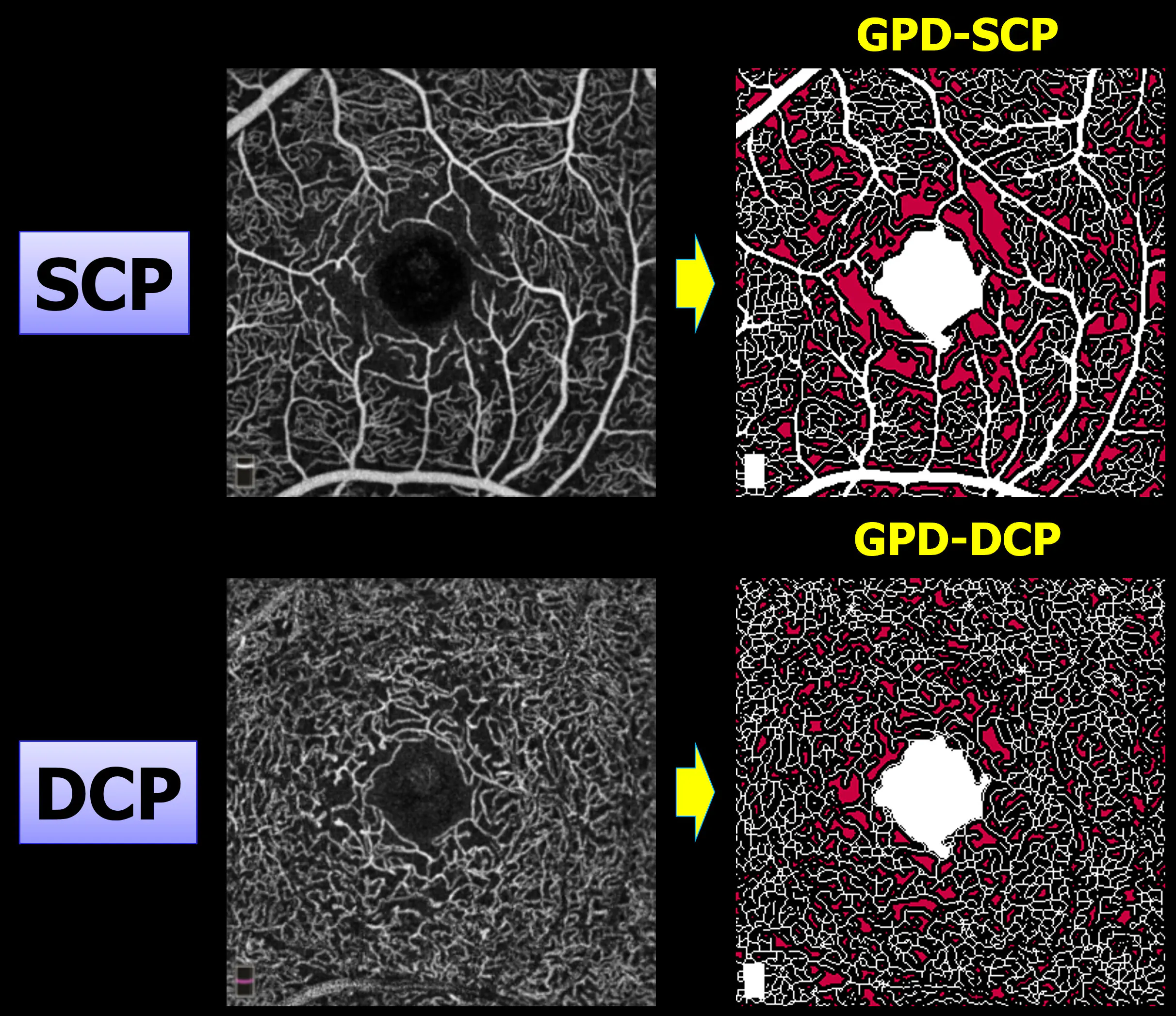
Figure 1. Representative en face OCTA images and corresponding GPD maps in the superficial and DCPs. GPD can be calculated in individual retinal capillary plexuses to quantify layer-specific ischemic burden.
RATE OF MICROVASCULAR PROGRESSION ON OCTA
The clinical value of a biomarker depends on whether it can identify eyes at risk for future worsening, rather than simply describe disease at a single point in time. We therefore pursued two complementary longitudinal questions:
- Can OCTA parameters detect short-term progression of microvascular dysfunction in DR?
- Can baseline GPD-DCP on OCTA predict clinically relevant complications?
To address the first question, we conducted a 1-year prospective study of 320 eyes (208 patients), evaluating longitudinal changes in multiple OCTA metrics at baseline, 6 months, and 12 months, stratified by DR severity: non-referable versus referable DR.14 In eyes with referable DR (moderate nonproliferative DR or worse), GPD-DCP showed significant worsening as early as 6 months, whereas no other OCTA metric showed a significant change.14 In eyes with nonreferable DR, no metric changed significantly at 6 months, and only vessel density in the superficial capillary plexus declined at 12 months. These findings demonstrate how OCTA can capture time-varying microvascular damage across different plexuses; moreover, GPD-DCP was more sensitive than other metrics to microvascular progression in eyes with referable DR.14
We next wanted to understand whether baseline GPD-DCP could predict progression of retinal nonperfusion beyond the macula. In a prospective longitudinal study, greater baseline GPD-DCP was independently associated with retinal nonperfusion progression on UWF-FA over 2 years. Although baseline UWF-FA nonperfusion remained the strongest predictor of subsequent progression, GPD-DCP provided independent prognostic information using macular OCTA scan. Thus, when serial FA is not practical or desirable, OCTA may help identify eyes at greater risk of progressive retinal nonperfusion.15
PREDICTING CLINICALLY RELEVANT COMPLICATIONS
We then investigated whether baseline GPD-DCP could predict the onset of clinically relevant DR complications, defined as worsening DR severity on UWF-FA, loss of vision, vitreous hemorrhage, initiation of anti-VEGF therapy, or need for panretinal photocoagulation.16 In a 2-year prospective study of 175 patients (265 eyes), 48 eyes (18.1%) from 41 patients (23.4%) experienced a complication. We found that higher baseline GPD-DCP was associated with an increased risk and earlier onset of complications.16 Each standard deviation increase in GPD-DCP was associated with a 77% higher hazard of complications and a 70-day reduction in complication-free survival. When eyes were dichotomized using a Youden’s J-derived threshold of 3.41%, those with GPD-DCP above this threshold developed complications approximately 4 months earlier than those below the threshold (Figure 2).16 These results suggest that GPD-DCP may serve as an objective metric for prognosticating and individualized care in DR.
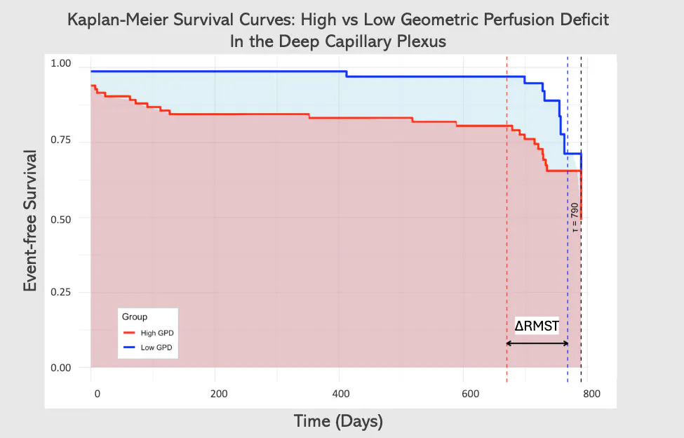
Figure 2. This Kaplan-Meier curve depicts the survival rates of eyes with high GPD-DCP versus eyes with low GPD-DCP. Event-free survival probability is illustrated on the vertical axis. Time in days is depicted on the horizontal axis. The dotted vertical lines represent the unadjusted restricted mean survival time (red for high GPD group, blue for low GPD group). The difference between the survival times is represented by the arrow labeled ∆RMST.
CAN OCTA PROVIDE THE ANSWER?
While OCTA cannot replace established methods of DR assessment, it offers a complementary advantage: noninvasive, layer-specific quantification of macular capillary ischemia. Across our studies, GPD-DCP is consistently associated with clinically important features of DR. Further validation is needed before GPD-DCP can be incorporated into routine clinical decision making. Additional studies are needed to calibrate this metric across different imaging devices, acquisition protocols, and diverse patient populations.
Nevertheless, current evidence suggests that OCTA-derived GPD-DCP may complement conventional DR grading, providing a quantitative, noninvasive measure of retinal ischemic burden.
1. Silva PS, Marcus DM, Liu D, et al. Association of ultra-widefield fluorescein angiography-identified retinal nonperfusion and the risk of diabetic retinopathy worsening over time. JAMA Ophthalmol. 2022;140(10):936-945. doi.org/10.1001/jamaophthalmol.2022.3130
2. Ong JX, Konopek N, Fukuyama H, Fawzi AA. Deep capillary nonperfusion on OCT angiography predicts complications in eyes with referable nonproliferative diabetic retinopathy. Ophthalmol Retina. 2023;7(1):14-23. doi:10.1016/j.oret.2022.06.018
3. Wykoff CC, Yu HJ, Avery RL, Ehlers JP, Tadayoni R, Sadda SR. Retinal non-perfusion in diabetic retinopathy. Eye (Lond). 2022;36(2):249-256. doi.org/10.1038/s41433-021-01649-0
4. de Barros Garcia JMB, Isaac DLC, Avila M. Diabetic retinopathy and OCT angiography: clinical findings and future perspectives. Int J Retina Vitreous. 2017;3:14. doi.org/10.1186/s40942-017-0062-2
5. Waheed NK, Rosen RB, Jia Y, et al. Optical coherence tomography angiography in diabetic retinopathy. Prog Retin Eye Res. 2023;97:101206. doi.org/10.1016/j.preteyeres.2023.101206
6. Couturier A, Mané V, Bonnin S, et al. Capillary plexus anomalies in diabetic retinopathy on optical coherence tomography angiography. Retina. 2015;35(11):2384-2391. doi.org/10.1097/iae.0000000000000859
7. Hwang TS, Gao SS, Liu L, et al. Automated quantification of capillary nonperfusion using optical coherence tomography angiography in diabetic retinopathy. JAMA Ophthalmol. 2016;134(4):367-373. doi.org/10.1001/jamaophthalmol.2015.5658
8. Zhang M, Hwang TS, Dongye C, Wilson DJ, Huang D, Jia Y. Automated quantification of nonperfusion in three retinal plexuses using projection-resolved optical coherence tomography angiography in diabetic retinopathy. Invest Ophthalmol Vis Sci. 2016;57(13):5101-5106. doi.org/10.1167/iovs.16-19776
9. Chen S, Moult EM, Zangwill LM, Weinreb RN, Fujimoto JG. Geometric perfusion deficits: a novel OCT angiography biomarker for diabetic retinopathy based on oxygen diffusion. Am J Ophthalmol. 2021;222:256-270. doi.org/10.1016/j.ajo.2020.09.007
10. Nesper PL, Ong JX, Fawzi AA. Deep capillary geometric perfusion deficits on OCT angiography detect clinically referable eyes with diabetic retinopathy. Ophthalmol Retina. 2022;6(12):1194-1205. doi.org/10.1016/j.oret.2022.05.028
11. Kakihara S, Zhuang K, Fawzi AA. Macular deep capillary plexus ischemia as a biomarker for identifying referable diabetic retinopathy. Invest Ophthalmol Vis Sci. 2025;66(14):45. doi.org/10.1167/iovs.66.14.45
12. Kakihara S, Busza A, Yamaguchi TC, Fawzi AA. Posterior retinal ischemia correlates with vision in patients with diabetes. Invest Ophthalmol Vis Sci. 2025;66(6):5. doi.org/10.1167/iovs.66.6.5
13. Kakihara S, Busza AM, Duffy BV, Zhuang K, AbdelSalam M, Fawzi AA. Macular optical coherence tomography angiography biomarkers predict regional retinal nonperfusion patterns on ultrawidefield angiography in diabetes. Am J Ophthalmol. 2026;287:198-207. doi.org/10.1016/j.ajo.2026.03.031
14. Kakihara S, Zhuang K, AbdelSalam M, Yamaguchi TC, Fawzi AA. The importance of matching optical coherence tomography angiography metrics to diabetic retinopathy severity for detecting progression. Invest Ophthalmol Vis Sci. 2025;66(11):49. doi.org/10.1167/iovs.66.11.49
15. Busza AM, Kakihara S, Zhuang K, Yamaguchi TC, Pearce E, Fawzi AA. Baseline nonperfusion and deep capillary plexus ischemia predict two-year retinal nonperfusion progression in diabetic retinopathy. Invest Ophthalmol Vis Sci. 2026;67(1):64. doi.org/10.1167/iovs.67.1.64
16. Zhuang K, Kakihara S, Busza A, Duffy B, AbdelSalam M, Fawzi AA. Baseline deep capillary plexus ischemia predicts earlier diabetic retinopathy complications than foveal avascular enlargement. Am J Ophthalmol. 2026:S0002-9394(26)00464-2. doi.org/10.1016/j.ajo.2026.08.014


