AT A GLANCE
- Pediatric patients undergoing examination under anesthesia can be imaged in the flying baby position.
- Scanning laser ophthalmoscopy can capture peripheral pigmentary changes, fibrovascular stalks, vitreous seeding, peripheral avascularity, and more.
- Ultrasound can be useful for detecting mass-like lesions associated with retinoblastoma and membranous echoes suggestive of persistent hyperplastic primary vitreous.
Pediatric ocular examinations can be challenging due to limited patient cooperation. Examination under anesthesia or sedation is often conducted to simplify the process. Retinal imaging also helps clinicians better understand the anatomy, pathophysiology, diagnosis, and management of retinal disorders.
Devices such as the Retcam (Natus) are available to document cases in which a pediatric patient is in a lying down position under anesthesia. Here, we describe pediatric cases that used a standard scanning laser ophthalmoscopy (SLO)-based device, the Mirante (Nidek), as a screening tool for capturing 163° widefield multimodal images in the flying baby position.
CASE 1
A 2-year-old girl presented with dimness of vision with nystagmus in each eye for 9 months. Dilated fundus examination of each eye revealed peripheral pigmentary changes sparing the posterior pole (Figure 1), suggestive of salt and pepper retinopathy. OCT of each eye showed a maintained foveal contour. The patient was advised to undergo screening for toxoplasmosis, rubella cytomegalovirus, herpes simplex, and human immunodeficiency virus and vitamin A supplementation after consulting with her pediatrician.
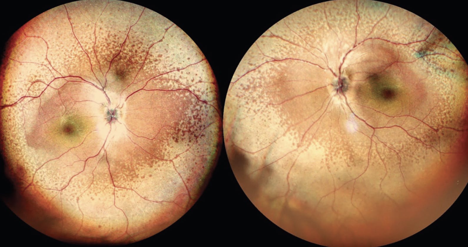
Figure 1. Widefield color imaging of each eye shows peripheral pigmentary changes sparing the posterior pole.
CASE 2
A 2.5-year-old boy presented with squinting in the left eye since birth. Dilated fundus examination revealed a fibrovascular stalk in the left eye extending from the disc to the posterior capsule of the lens suggestive of persistent hyperplastic primary vitreous (Figure 2). OCT showed an altered foveal contour. Ultrasound of the left eye showed membranous echoes with low to moderate spikes and restricted movements suggestive of persistent hyperplastic primary vitreous. The patient was advised to undergo refraction, use low vision aids, and follow up every 6 months.
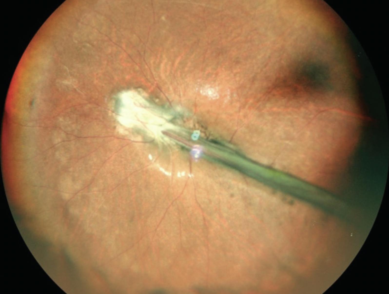
Figure 2. Widefield imaging of the left eye shows a fibrovascular stalk extending from the optic disc to the posterior capsule of the lens.
CASE 3
A 5-year-old boy presented with dimness of vision in the left eye since birth. Dilated fundus examination revealed a large optic disc with glial tissue, straightening of the vessels, and pigmentation, suggestive of morning glory syndrome. OCT showed subretinal fluid suggestive of a retinal detachment associated with morning glory syndrome (Figure 3). No vitreoretinal intervention was recommended, and the patient was scheduled for follow up every 6 months.
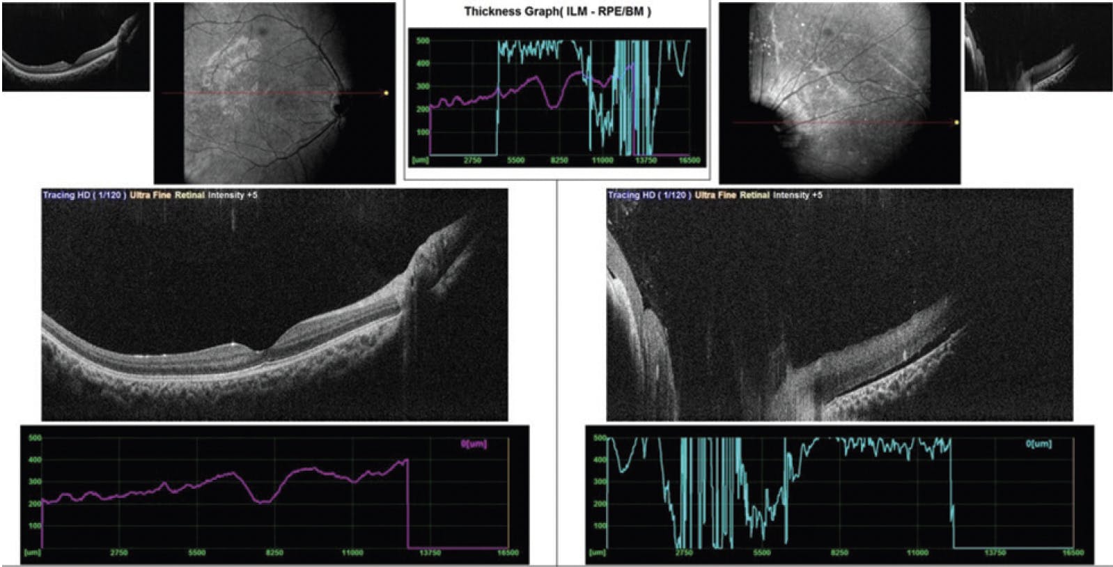
Figure 3. OCT of the left eye shows altered foveal contour with subretinal fluid suggestive of a retinal detachment associated with morning glory syndrome.
CASE 4
A 2.5-year-old girl presented with dim vision and whitish reflex in the right eye for 2 months. Dilated fundus examination of the right eye revealed multiple whitish nodular masses with vitreous seeding, indicative of retinoblastoma (Figure 4). Ultrasound of the right eye revealed hyperechoic homogenous masses with high spikes and restricted movements suggestive of calcification associated with retinoblastoma. The patient underwent enucleation of the right eye.
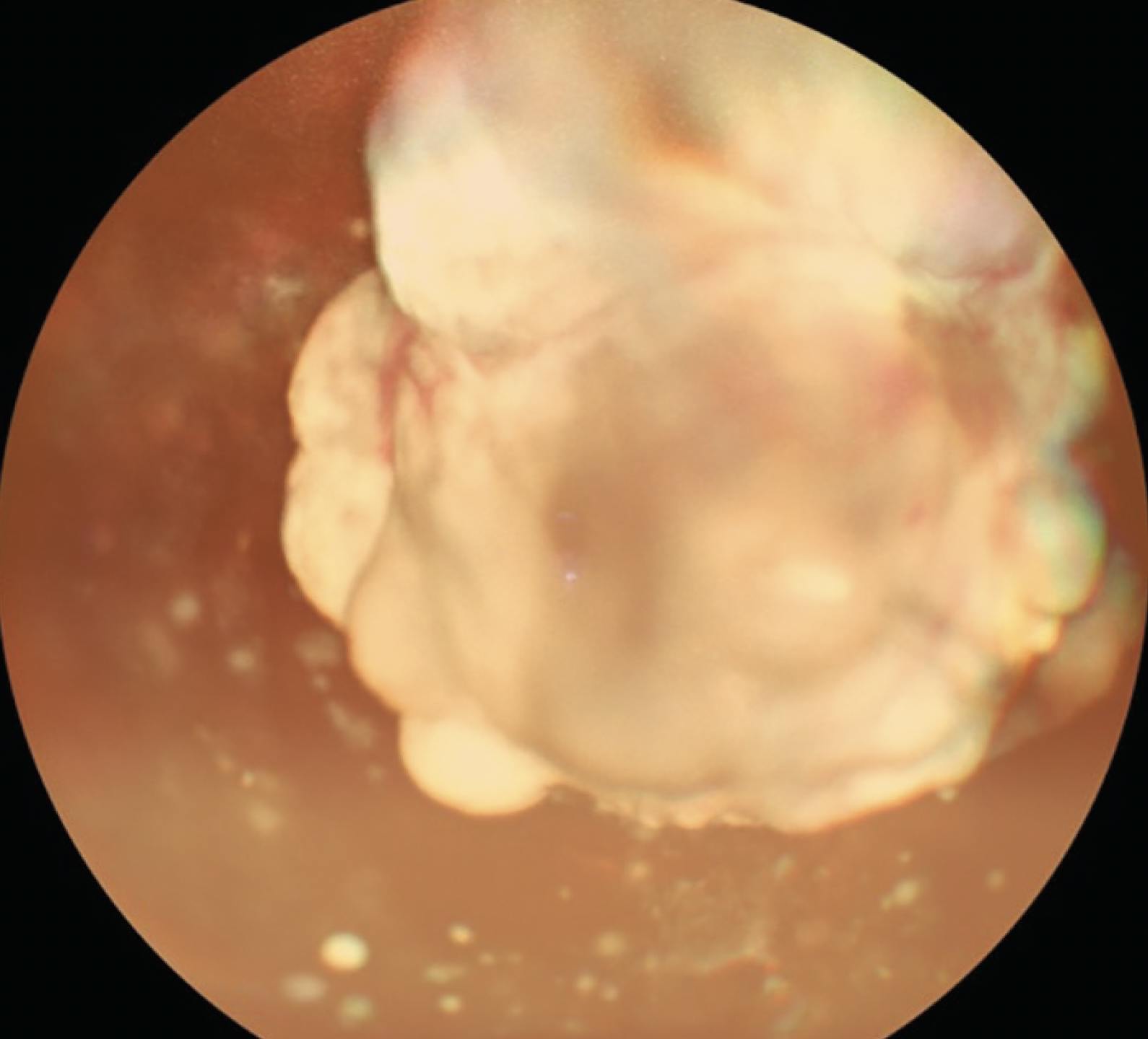
Figure 4. Widefield color imaging of the right eye shows multiple whitish nodular masses with vitreous seeding associated with retinoblastoma.
CASE 5
A 5-year-old boy presented with dimness of vision with squinting in the right eye for 1 year. Dilated fundus examination of the right eye revealed a falciform fold extending temporally from the disc with straightening of the vessels with peripheral avascularity (Figure 5). The left eye had flat traction temporally with straightening of the vessels with peripheral avascularity inferiorly. OCT of each eye showed altered foveal contour. No vitreoretinal intervention was recommended, and the patient was scheduled for follow up every 3 months.
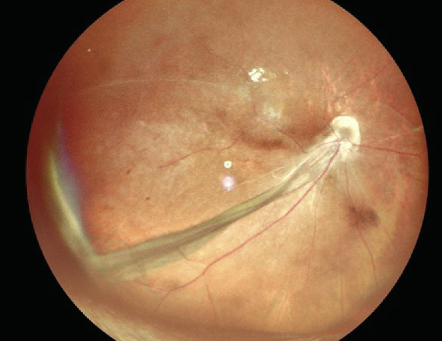
Figure 5. Widefield color imaging of the right eye reveals falciform extending temporally from the disc with straightening of the vessels with peripheral avascularity.
CASE 6
A 6-year-old boy presented with outward deviation of the left eye and dimness of vision (more at night) in each eye since he was 2.5 years of age. BCVA was 6/60 OU. Dilated fundus examination of each eye revealed a granular fundus suggestive of heredomacular degeneration. OCT of each eye showed altered foveal contour with thinning. Fundus autofluorescence showed hypoautofluorescence with a ring of hyperautofluorescence in each eye, suggestive of heredomacular degeneration (Figure 6). The patient was advised to undergo pattern electroretinography and visual evoked potential testing but was lost to follow-up.
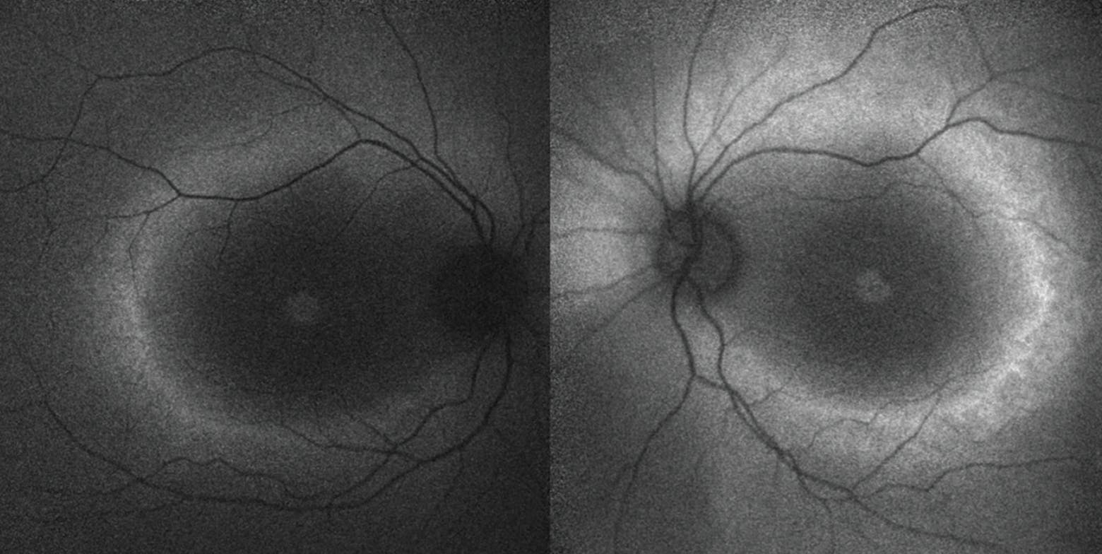
Figure 6. Fundus autofluorescence of each eye shows hypoautofluorescence with a ring of hyperautofluorescence, suggestive of heredomacular degeneration.
CASE 7
A 1.5-year-old boy, born prematurely at 7.5 month’s gestation, presented with dimness of vision in each eye since birth. The patient had received anti-VEGF injections for stage 4 retinopathy of prematurity elsewhere. Dilated fundus examination of each eye showed fibrovascular proliferation at the disc with large areas of avascularization in the left eye. OCT of each eye showed an altered foveal contour (Figure 7). The patient underwent barrage laser treatment to the avascular zones.

CASE 8
A 9-month-old boy presented with leukocoria in each eye since he was 2 months of age. Dilated fundus examination of each eye revealed multiple white lesions at the posterior pole with venous tortuosity and prominence involving the disc and macula, likely suggesting retinoblastoma. Ultrasound of each eye showed heterogenous mass-like lesions with moderate to high spikes suggestive of calcification associated with retinoblastoma (Figure 8). The patient was referred to an ocular oncologist.


_1773249222.png?auto=compress,format&w=75)


