
KEY TAKEAWAYS
- Uveal melanoma (UM) is the most common primary intraocular cancer in adults, with an annual incidence of 5.1 cases per 1 million individuals in the United States.
- The three strongest predictors of transformation of a choroidal nevus into UM were found to be tumor thickness > 2 mm on ultrasonography, subretinal fluid on OCT, and orange pigment on fundus autofluorescence.
- The risk for transformation of a nevus into UM increases sharply with age, becoming nearly 75-times more likely by the eighth decade of life; still, careful monitoring must be guided by objective criteria rather than age alone.
Uveal melanoma (UM) is the most common primary intraocular cancer in adults, with an annual incidence of 5.1 cases per 1 million individuals in the United States and a propensity to affect individuals with more lightly pigmented skin.1,2 UM mainly presents in the choroid (90%), with fewer tumors arising in the ciliary body (6%) and iris (4%).3,4 Prognosis worsens when the ciliary body is involved, owing to the greater tumor thickness, greater risk for cytogenetic abnormalities, and higher metastatic potential.5 In one series of 1,059 patients tested for genetic abnormalities, ciliary body tumors demonstrated the highest odds ratio (OR) for chromosome 3 monosomy (OR = 8.17, P < .001) and chromosome 8q gain (OR = 102.87, P = .001), two alterations that are strongly predictive of metastasis.5
Despite advances in local control of UM, approximately 50% of patients develop metastasis, mainly to the liver via hematogenous spread.4,6,7 This persistent risk for systemic spread underscores the need for early detection and structured, risk-based monitoring to improve long-term outcomes. While risk-assessment tools have been published and apply across all ages, nevus transformation into melanoma remains rare in patients under 30 years of age.8-10
In this article, we describe a case of melanocytic growth of a choroidal nevus in a 28-year-old patient, our treatment approach, and the importance of balancing vigilance with measured follow-up in patients of all ages.
RISK FACTORS FOR CHOROIDAL NEVUS TRANSFORMATION INTO MELANOMA
Careful monitoring must be guided by objective criteria rather than age alone. One widely used framework to standardize the criteria for nevus transformation to melanoma is the mnemonic “To Find Small Ocular Melanoma Doing Imaging” (ie, TFSOM-DIM):11
- Thickness > 2 mm (by ultrasonography)
- Fluid, subretinal (by OCT)
- Symptoms of VA loss to ≤ 20/50 (by Snellen acuity)
- Orange pigment (by fundus autofluorescence [FAF])
- Melanoma showing acoustic hollowness
- (by ultrasonography)
- DIaMeter > 5 mm (by fundus photography)
Previous analyses of 1,329 and 1,287 patients with small melanocytic choroidal tumors first highlighted these important features as key predictors of growth and occasional metastasis, with risks compounding when several of these factors were present.12,13 A larger subsequent series of 3,806 patients with choroidal nevus showed that the probability of transformation into melanoma increased when more risk factors were present at the time of diagnosis and over time.11 The 5-year Kaplan-Meier transformation rates were 1% with no factors, 11% with any one factor, 22% with two factors, 34% with three factors, and > 50% with at least four factors.11 The three strongest predictors of transformation of nevus into melanoma included tumor thickness > 2 mm on ultrasonography, subretinal fluid on OCT, and orange pigment on FAF. A smaller effect was noted for symptoms of visual acuity loss on Snellen acuity, melanoma acoustic hollowness on ultrasonography, and tumor diameter > 5 mm on fundus photography.11
In addition to TFSOM-DIM, the MOLES scoring system—Mushroom shape on ultrasonography, Orange pigment on fundus photography and FAF, Large size (≥ 3 disc diameters in base or thickness ≥ 1 mm), Enlarging tumor with growth, and Subretinal fluid on OCT—has been proposed for use by nonspecialists and communities with limited access to advanced imaging techniques in an effort to guide monitoring and referral.14 Despite its good specificity and sensitivity, the accuracy of the MOLES system diminishes for very small tumors.14 These predictive algorithms can help clinicians implement targeted surveillance and timely intervention for patients with one or more high-risk features.
CASE REPORT
A 28-year-old White man with no ocular melanocytosis was observed with a choroidal nevus in his right eye for 6 years. Initial fundoscopic imaging elsewhere showed a flat pigmented lesion measuring 4 mm x 4 mm in diameter (Figure 1A, arrow) and demonstrated hypoautofluorescence (Figure 1B, arrow). He was referred to our team with a possible tumor growth of a nevus into melanoma. His BCVA was 20/25 OU. Fundus examination of the right eye confirmed a dome-shaped pigmented mass in the nasal juxtapapillary region (Figure 2A) with overlying orange pigment on FAF (Figure 2B) and ultrasonographic acoustic hollowness (Figure 2C). OCT showed overlying subretinal fluid and a pocket of subretinal fluid inferonasally (Figure 2D, arrow). The lesion measured 9 mm in largest basal diameter and 2.6 mm in thickness. The fellow eye was unremarkable. By TFSOM-DIM criteria, the tumor met four of six risk factors, and the patient was subsequently diagnosed with a small, growing choroidal melanoma. Plaque radiotherapy with apex dose of 7,000 cGy was subsequently provided.
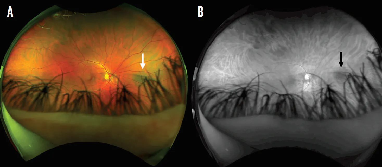
Figure 1. Fundus photography (A) and FAF (B) showed a flat pigmented choroidal nevus with hypoautofluorescence (arrows) and no orange pigment.
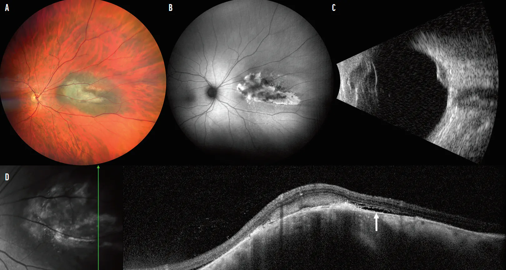
Figure 2. Fundus photography at presentation to our clinic demonstrated interval growth of the lesion (A). FAF showed hyperautofluorescence and orange pigment and dependent subretinal fluid (B). B-scan ultrasonography showed an echolucent choroidal mass of 9 mm in largest basal diameter and 2.6 mm in thickness (C). OCT vertical cut through the lesion showed shallow subretinal fluid, including a pocket of fluid superiorly and tracking down inferiorly (D).
AGE-STRATIFIED TRANSFORMATION RISK
The likelihood of nevus transformation increases with advancing age based on a reliable study by Singh et al.8,11,15 This study evaluated a population-based model of the US White population and found that the risk is negligible in teenagers (one in 270,000) but increases sharply with age, becoming nearly 75-times more likely by the eighth decade of life (one in 3,600).8 Age also influences the anatomic distribution of tumors, as shown in our series of 8,033 patients, with the proportion of iris and ciliary body involvement more predominant in younger patients (29%, P < .05) compared with older adults (9%, P < .05), although the choroid remained the most common site for UM for all age brackets.4
Long-term metastasis rates in this same group were markedly age dependent as well, with patients older than 60 years of age experiencing a higher proportion of metastasis and death compared with younger groups.4 The prognostic effect of age is even more pronounced with multiple TFSOM-DIM features.11 These findings should prompt more frequent imaging and longer-term follow-up in older patients with high-risk features. While transformation is exceedingly uncommon in teenagers and children, the small subset who develop melanoma may still carry a risk of metastasis and mortality.9,10,15,16
PEDIATRIC UVEAL MELANOMA
Pediatric UM, although uncommon, presents unique physiological and clinical challenges distinct from adult UM. A pooled study sample of 5,533 children with 48 choroidal nevi demonstrated that the prevalence of choroidal nevi increased steadily with age.15 Longitudinal data was available for 23 of these nevi and showed that 78% remained stable, a minority (22%) demonstrated subtle growth, and none transformed into melanoma.15 While this study underscores the rarity of choroidal nevus transformation to melanoma in pediatric populations, the occurrence of pediatric UM is well documented.10 In a multicenter series of 133 pediatric patients ≤ 12 years of age, pediatric UM most often arose in the choroid or ciliary body and less commonly in the iris, with ocular melanocytosis (a predisposing factor for the development of UM) present in 15% of choroidal/ciliary body tumors and 11% of iris tumors, compared with 0.04% in the general population.10 These findings align with our own observation in children and teenagers, where most tumors have originated in the choroid, with lower rates in the iris and ciliary body.9
In our series, metastasis rates increased over decades of follow-up, and treatment for pediatric UM most often included radiotherapy or enucleation.9 At the more severe end of the spectrum, a small series of 13 infants under 2 years of age found that all eyes were surgically removed (85% through enucleation and 15% through exenteration), with around 15% of patients developing metastasis and 8% dying within 6 months.16 Although rare, pediatric UM can follow an aggressive course; outcomes may be severe, emphasizing the need for clinical vigilance.
RARE, BUT POSSIBLE
Rare transformation of choroidal nevus into melanoma can occur, such as in the young adult discussed here. Use of the structured evaluation with TFSOM-DIM, combined with careful documentation of growth and risk features, remains a cornerstone of early melanoma detection.
Acknowledgement: Support provided in part by the Jerry A. Shields, MD, Eye Cancer Fund, Philadelphia, PA (CLS) and the Eye Tumor Research Foundation, Philadelphia, PA (CLS). The funders had no role in the design and conduct of the study, in the collection, analysis, and interpretation of the data, and in the preparation, review, or approval of the manuscript. Carol L. Shields, MD, has had full access to all the data in the study and takes responsibility for the integrity of the data.
1. Singh AD, Turell ME, Topham AK. Uveal melanoma: Trends in incidence, treatment, and survival. Ophthalmology. 2011;118(9):1881-1885.
2. Negretti GS, Bayasi F, Goldstein S, et al. Association of Fitzpatrick skin type with metastatic risk from uveal melanoma in 854 consecutive patients at a single center. Eye (Lond). 2024;38(3):565-571.
3. Shields CL, Furuta M, Thangappan A, et al. Metastasis of uveal melanoma millimeter-by-millimeter in 8033 consecutive eyes. Arch Ophthalmol. 2009;127(8):989-998.
4. Shields CL, Kaliki S, Furuta M, et al. Clinical spectrum and prognosis of uveal melanoma based on age at presentation in 8033 cases. Retina. 2012;32(7):1363-1372.
5. Shields CL, Say EA, Hasanreisoglu M, et al. Cytogenetic abnormalities in uveal melanoma based on tumor features and size in 1059 patients: The 2016 W. Richard Green Lecture. Ophthalmology. 2017;124(5):609-618.
6. Bansal R, Sener H, Ganguly A, et al. Metastasis-free survival of uveal melanoma by tumour size category based on The Cancer Genome Atlas classification in 1001 cases. Clin Exp Ophthalmol. 2025;53(2):175-183.
7. Diener-West M, Reynolds SM, Agugliaro DJ, et al. Development of metastatic disease after enrollment in the COMS trials for treatment of choroidal melanoma: Collaborative Ocular Melanoma Study Group Report No. 26. Arch Ophthalmol. 2005;123(12):1639-1643.
8. Singh AD, Kalyani P, Topham A. Estimating the risk of malignant transformation of a choroidal nevus. Ophthalmology. 2005;112(10):1784-1789.
9. Shields CL, Kaliki S, Arepalli S, et al. Uveal melanoma in children and teenagers. Saudi J Ophthalmol. 2013;27(3):197-201.
10. Masoomian B, Dalvin LA, Riazi-Esfahani H, et al. Pediatric ocular melanoma: A collaborative multicenter study and meta-analysis. J AAPOS. 2023;27(6):316-324.
11. Shields CL, Dalvin LA, Ancona-Lezama D, et al. Choroidal nevus imaging features in 3806 cases and risk factors for transformation into melanoma in 2355 cases: The 2020 Taylor R. Smith and Victor T. Curtin Lecture. Retina. 2019;39(10):1840-1851.
12. Shields CL, Shields JA, Kiratli H, et al. Risk factors for growth and metastasis of small choroidal melanocytic lesions. Ophthalmology. 1995;102(9):1351-1361.
13. Shields CL, Cater J, Shields JA, et al. Combination of clinical factors predictive of growth of small choroidal melanocytic tumors. Arch Ophthalmol. 2000;118(3):360-364.
14. Roelofs KA, O’Day R, Al Harby L, et al. The MOLES system for planning management of melanocytic choroidal tumors: is it safe? Cancers (Basel). 2020;12(5):1311.
15. Raval V, Bellerive C, Singh AD, et al. Choroidal nevi in children: Prevalence, age of onset, and progression. J AAPOS. 2021;25(4):225.e1-225.e6.
16. Yousef YA, Alkilany M. Characterization, treatment, and outcome of uveal melanoma in the first two years of life. Hematol Oncol Stem Cell Ther. 2015;8(1):1-5.