KEY TAKEAWAYS
- Vitreoretinal surgeons have used fibrin glue off-label as a localized hemostatic agent in the setting of trauma, iatrogenic retinal breaks, and diabetic neovascularization, and as a form of retinopexy/tamponade to treat rhegmatogenous retinal detachment.
- When using fibrin glue intraocularly, the retinal surface must be carefully dried under air. The thick component of the glue is applied first, followed by the thin component shortly after.
- Potential complications with the use of fibrin glue include displacement of the glue posteriorly, subretinal migration, and epiretinal membrane or proliferative vitreoretinopathy formation because fibrin can act as a scaffold.
Fibrin glue, a biologic tissue adhesive that recreates the final step of the coagulation cascade, has been a common tool in ophthalmic surgery for decades. It consists of fibrinogen and thrombin, and when the two components are combined, they quickly polymerize into a fibrin clot. That clot forms a temporary scaffold that adheres tissues together.1 Fibrin glue is often used in anterior segment surgery, such as conjunctival closure in strabismus surgery, conjunctival autografts in pterygium surgery, sealing wounds and perforations in corneal surgery, and amniotic membrane transplantation for ocular surface disease.2-6
The use of fibrin glue in vitreoretinal surgery is more recent and still evolving. Surgeons have used it as a localized hemostatic agent in the setting of trauma, iatrogenic retinal breaks, and diabetic neovascularization, and as a form of retinopexy/tamponade to treat rhegmatogenous retinal detachment (RRD).7-13
Here, we share the surgical steps and considerations for using this tool in your retina OR (Video).
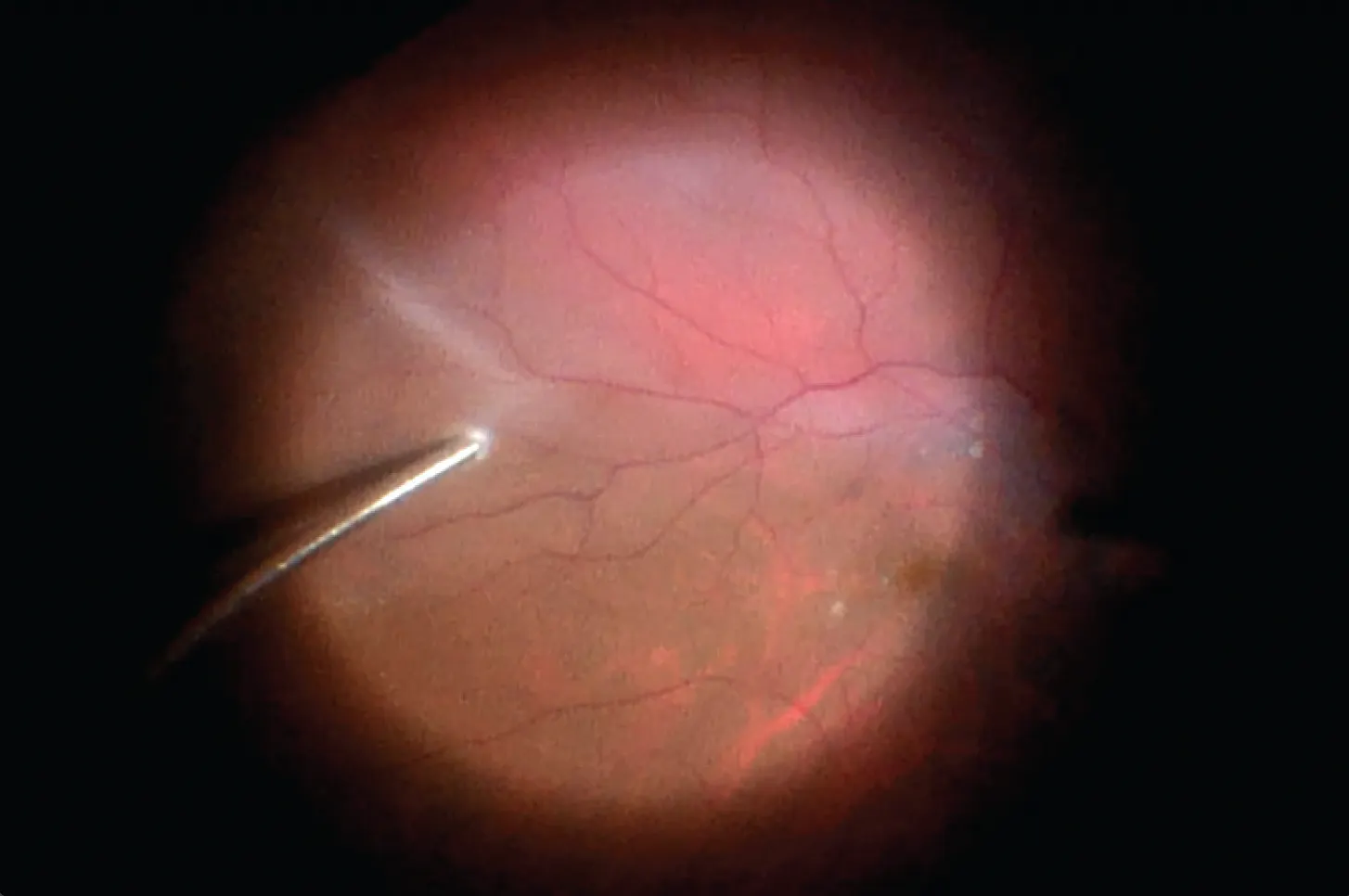
Video. Fibrin Glue-Assisted RD Repair
THE PROBLEM WITH TRADITIONAL TAMPONADES
Tamponade agents such as gas (eg, air, SF6, C3F8), silicone oil, and perfluoro-n-octane (PFO) are effective, but they come with several constraints and potential complications. They often require postoperative positioning, can significantly reduce vision for weeks or months, prevent patients from flying or traveling to high elevations, carry risks such as elevated IOP, and, in the case of silicone oil, require a second surgery for removal.14,15
WHEN TO CONSIDER GLUE
Using fibrin glue shifts the strategy from filling the eye to mechanically seal a break toward directly sealing the break itself without the need for tamponade. Using this strategy, the eye could be filled with balanced salt solution at the end of the case, resulting in clearer postoperative vision—which is particularly helpful for monocular patients—and allowing for air travel or driving to high elevations. In addition, fibrin glue self-dissolves in approximately 1 to 2 weeks, which is enough time for the chorioretinal adhesion from laser to form.
Patients with RRD who may benefit from fibrin glue include those who are poor candidates for gas tamponade, such as those who are monocular, unable to tolerate postoperative positioning, or who must travel by air or to high elevations. Others who may do well with fibrin glue include those with RRDs involving inferior retinal breaks and complex proliferative vitreoretinopathy requiring large retinectomies or giant retinal tears, where stabilization of the retinal edges is needed to prevent slippage (Figure 1).
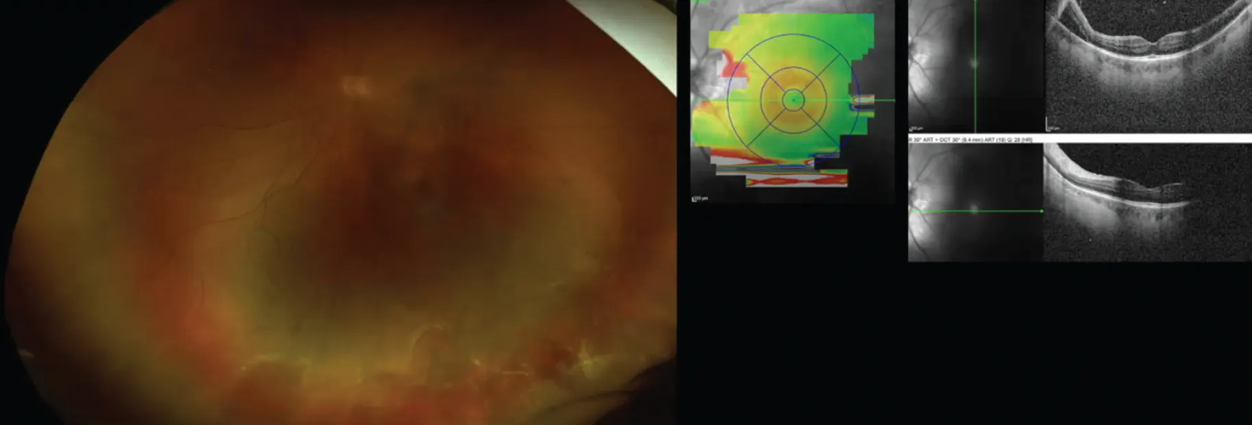
Figure 1. A 41-year-old man presented with a persistent inferior RD after scleral buckling surgery. In lieu of a traditional long-lasting tamponade, the surgeon applied fibrin glue.
Patients who can benefit from fibrin glue’s hemostatic properties include those with uncontrolled bleeding from neovascularization (eg, proliferative diabetic retinopathy/tractional RDs) and bleeding from trauma, anticoagulation, or intraoperative iatrogenic causes.
Fibrin glue has also been successfully used to assist with macular hole closure and optic disc pit-related macular detachments.7,11,16
THE SURGICAL STEPS
The early steps are similar to a standard RD repair: core vitrectomy, induction of posterior vitreous detachment (if needed), meticulous peripheral shaving, flattening of the retina with fluid-air exchange or drainage through the break or a retinotomy, and endolaser around the break(s).
The retinal surface must be carefully dried under air, which is an important condition for the fibrin glue to work. The thick component of the glue is applied first, followed by the thin component shortly after (Figure 2). It is important to avoid using excessive amounts of glue. Only a drop at a time is necessary for each of the thick and thin components, and you can always apply more if needed. The polymerization occurs within seconds and is evident when the glue turns opaque.
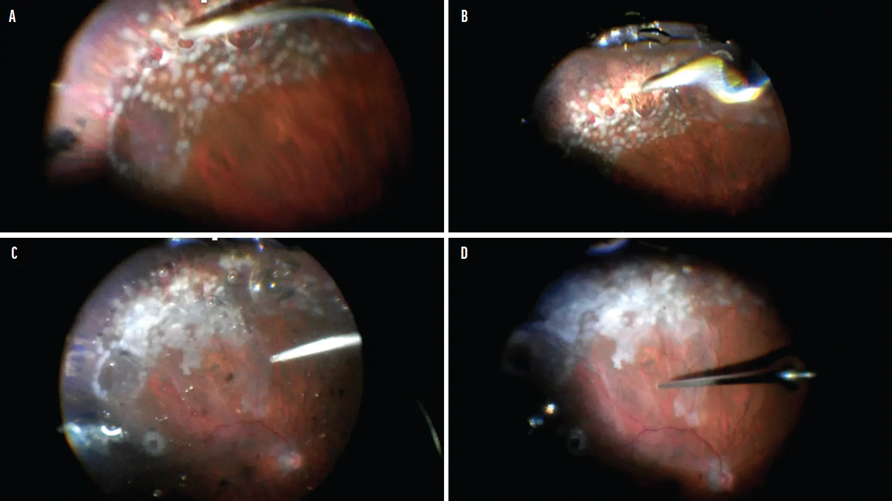
Figure 2. When applying fibrin blue, surgeons must instill the thick component (A) followed by the thin component (B). In this patient’s case, the retina remained reattached when tested under fluid (C), and fluid-air exchange was repeated (D).
The eye may need to be tilted toward the target area to minimize the amount of glue migrating posteriorly to the disc and macula. Sometimes, forceps can be used to pick up the partially formed coagulum from the posterior pole and move it back to the target area.
PITFALLS OF FIBRIN GLUE IN THE RETINA
The techniques described here are off-label applications of fibrin glue. Potential complications include displacement of the glue posteriorly, subretinal migration, and epiretinal membrane or proliferative vitreoretinopathy formation because fibrin can act as a scaffold. Intraocular inflammation is also possible because fibrin glue is a biologic product. There is a theoretical risk of viral or bacterial transmission because the glue is derived from pooled plasma, although modern processing techniques minimize this risk.17
USE WITH CARE
Fibrin glue’s ability to provide localized adhesion, hemostasis, and temporary support without the limitations of gas or oil make it an appealing option in select cases. Used judiciously, this tool, already a mainstay in most ORs, can be a valuable adjunct in the retina OR, offering flexibility when traditional tamponades are infeasible or less desirable.
1. Levicheva EA, Linkova DD, Farafontova EA, et al. Fibrin glues: proteins, mechanism of action, classification, and application. Int J Mol Sci. 2025;27(1):447.
2. Yang MB, Melia M, Lambert SR, Chiang MF, Simpson JL, Buffenn AN. Fibrin glue for closure of conjunctival incision in strabismus surgery: a report by the American Academy of Ophthalmology. Ophthalmology. 2013;120(9):1935-1940.
3. Koranyi G, Seregard S, Kopp ED. Cut and paste: a no suture, small incision approach to pterygium surgery. Br J Ophthalmol. 2004;88(7):911-914.
4. Anderson NJ, Hardten DR. Fibrin glue for the prevention of epithelial ingrowth after laser in situ keratomileusis. J Cataract Refract Surg. 2003;29(7):1425-1429.
5. Kaufman HE, Insler MS, Ibrahim-Elzembely HA, Kaufman SC. Human fibrin tissue adhesive for sutureless lamellar keratoplasty and scleral patch adhesion a pilot study. Ophthalmology. 2003;110(11):2168-2172.
6. Duarte MC, Kim T. Sutureless lamellar keratoplasty: a modified approach for fibrin glue application. Cornea. 2007;26(9):1127-1128.
7. Tyagi M, Basu S. Glue-assisted retinopexy for rhegmatogenous retinal detachments (GuARD): A novel surgical technique for closing retinal breaks. Indian J Ophthalmol. 2019;67(5):677-680.
8. Desai A, Bhopalka AK, Tyagi M, Sharma SV, Takkar B. Glue-assisted retinopexy for retinal detachments study report 2: Fibrin glue as a surgical adjunct in complex rhegmatogenous retinal detachment. J Vitreoretin Dis. 2024;8(4):435-441.
9. Wang Q, Zhao J, Xu Q, Han C, Hou B. Intraocular application of fibrin glue as an adjunct to pars plana vitrectomy for rhegmatogenous retinal detachment. Retina. 2020;40(4):718-724.
10. Takkar B, Ketkar MR, Narula R, Tyagi M, Dave VP, Narayanan R. A pilot randomized controlled trial evaluating hemostasis with fibrin glue during surgery for proliferative diabetic retinopathy. Am J Ophthalmol. 2025;272:117-125.
11. Hansraj S, Mb T, Takkar B, et al. Clinical outcomes of fibrin glue assisted hemostasis in persistent intraoperative optic disc bleeding. Int J Retina Vitreous. 2026;12(1):33.
12. Aydin E, Eris E, Kazanci L, Uyar OM. Reattachment of rhegmatogenous retinal detachment via fibrin tissue adhesive. Korean J Ophthalmol. 2021;35(3):173-178.
13. Stuart A. New Approaches to Securing Retinal Breaks During Surgery. EyeNet. November 1, 2022. Accessed March 11, 2026. www.aao.org/eyenet/article/new-approaches-to-securing-retinal-breaks
14. Valentín-Bravo FJ, García-Onrubia L, Andrés-Iglesias C, et al. Complications associated with the use of silicone oil in vitreoretinal surgery: A systemic review and meta-analysis. Acta Ophthalmol. 2022;100(4):e864-e880.
15. Vaziri K, Schwartz SG, Kishor KS, Flynn HW Jr. Tamponade in the surgical management of retinal detachment. Clin Ophthalmol. 2016;10:471-476.
16. Soni A, Singh SR, Chhablani J. Fibrin glue for treatment of optic disc maculopathy. Saudi J Ophthalmol. 2021;34(3):227-229.
17. Anshuman K, Ratra D. Commentary: Fibrin glue in rhegmatogenous retinal detachment repair-Are we there yet? Indian J Ophthalmol. 2019;67(5):683-684.