
KEY TAKEAWAYS
- Photobiomodulation (PBM) has been applied across many medical fields, including dermatology, neurology, and dentistry, where it has been shown to improve tissue repair, reduce neuropathic pain, and enhance bone remodeling.
- Ideal candidates for PBM include patients with intermediate dry AMD who are motivated to pursue a noninvasive treatment option.
- Patients typically undergo an initial series of nine treatment sessions over 3 to 5 weeks, followed by maintenance treatments every 4 to 6 months based on clinical response and patient preference.
- While PBM may improve visual function and slow disease progression, it is not curative. Outcomes may vary, and treatment benefits are generally more pronounced in earlier stages of disease.
The prevalence of AMD is projected to increase from approximately 200 million to more than 300 million individuals worldwide as global life expectancy rises.1,2 Accordingly, the development of therapies that can prevent, slow, or treat AMD has become critical (Figure). The treatment landscape for AMD has evolved substantially, from AREDS supplementation to anti-VEGF therapies and complement inhibitors, and, most recently, photobiomodulation (PBM). Currently, PBM offers a noninvasive therapeutic option for patients with dry AMD.
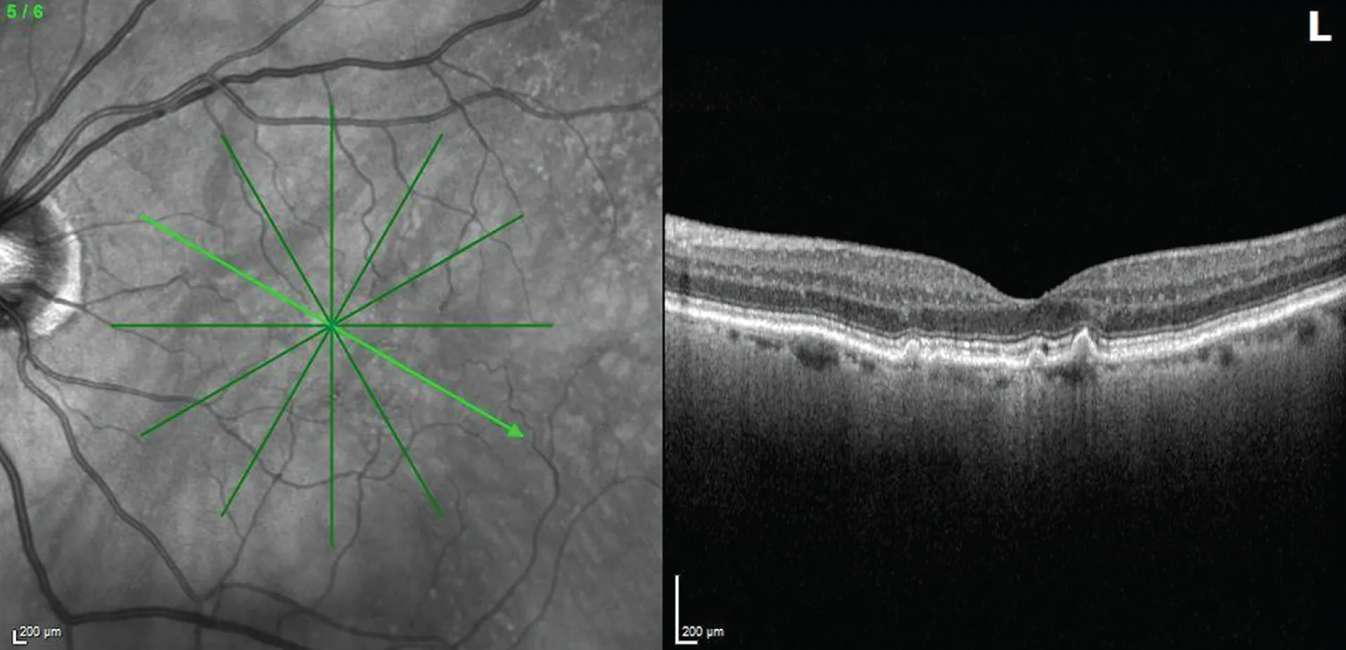
Figure. Patients such as this 68-year-old woman who has intermediate AMD, with the OCT of the left eye demonstrating drusen, may benefit from early treatment.
In this article, we review its mechanisms of action, current clinical evidence, and practical considerations for real-world implementation.
WHAT IS PHOTOBIOMODULATION?
PBM involves the application of low-level light therapy using low-power lasers or light-emitting diodes at specific wavelengths, typically ranging from 590 nm to 850 nm. PBM is a nonthermal, noninvasive therapy that is thought to act primarily at the mitochondrial level, enhancing cytochrome c oxidase activity and promoting ATP production.3 This aims to reduce oxidative stress and modulate inflammatory signaling pathways, ultimately promoting cellular survival and function.3
The biologic effects of PBM were first described in the 1960s, when researchers demonstrated its role in promoting wound healing.4 Since then, PBM has been applied across many medical fields, including dermatology, neurology, and dentistry, where it has been shown to improve tissue repair, reduce neuropathic pain, and enhance bone remodeling.3,5,6
The retina represents an ideal target for PBM due to its high metabolic demand and high mitochondrial numbers within photoreceptors, retinal pigment epithelial (RPE) cells, and ganglion cells. By enhancing mitochondrial function and reducing inflammatory mediators, PBM may modify key pathogenic pathways implicated in AMD, including oxidative stress, mitochondrial dysfunction, and chronic inflammation.7 Thus, PBM has emerged as a promising treatment to potentially alter the inflammatory and mitochondrial damage cycle of AMD and slow disease progression.
In the United States, the only FDA-approved PBM device for dry AMD is the Valeda Light Delivery System (Alcon). The system uses three wavelengths, yellow (590 nm), red (660 nm), and near-infrared (850 nm), to target multiple mitochondrial chromophores. In the European Union, both the Valeda system and the Eye-light (Espansione Group) have received CE marks for dry AMD. The Eye-light device, originally approved for dry eye disease and blepharitis, uses yellow (590 nm) and red (660 nm) wavelengths.
CLINICAL EVIDENCE
The LIGHTSITE I and II trials were randomized controlled studies evaluating the safety and efficacy of PBM using the Valeda system in patients with dry AMD. These studies demonstrated improvements in contrast sensitivity, BCVA, and stabilization of drusen volume compared with sham-treated controls. Treatment effects were more pronounced in patients with intermediate AMD compared with advanced disease.8,9
The LIGHTSITE III trial, a phase 3 multicenter, double-masked, prospective study, evaluated 100 patients with early to intermediate AMD over a 24-month period. Patients received nine treatment sessions (three times weekly) every 4 months.10 The study demonstrated a statistically significant and sustained improvement in BCVA, with a mean gain of 5.9 letters at month 24 compared with sham (P < .0001). In addition, the treatment group showed stabilization of drusen volume, whereas the sham group exhibited a trend toward progression. A lower incidence of new-onset geographic atrophy (GA; 7% vs 24%) and improvements in vision-related quality of life were also observed. Importantly, no phototoxicity or major safety concerns were identified.10
LIGHTSITE IIIB, an extension study, followed a subset of patients for an additional 13 months.11 There was a 20-month interruption period before the start of the extension study. During this interruption period, previously treated patients experienced a mean decline of 2.2 letters but regained approximately 5 letters after resuming therapy, with sustained benefit throughout the extension period.11 In contrast, patients originally assigned to the sham group experienced greater visual decline and limited recovery after initiating treatment. At approximately 4.5 years of follow-up since the initiation of LIGHTSITE III, a 9.9-letter difference was observed between the original treatment and sham groups. Subgroup analysis suggested greater benefit in patients with worse baseline visual acuity (worse than 70 ETDRS letters). No significant safety signals were identified, although three patients developed wet AMD during follow-up.
A recent randomized multicenter European study evaluated the Eye-light device in 152 eyes with dry AMD.7 Patients underwent two treatment sessions per week for 4 weeks. At the conclusion of treatment, a significantly higher proportion of treated eyes gained ≥ 5 letters compared with sham (20.3% vs 9%). Additionally, drusen volume remained stable in the treatment group compared with progression in the control group. The therapy was well tolerated, with no major adverse events reported.7
Limitations of Current Evidence
Despite promising results, limitations remain. The sample sizes in randomized controlled trials are relatively small, which may limit generalizability and increase susceptibility to confounding variables. Long-term data on progression to GA or wet AMD are limited. Furthermore, direct comparisons with established interventions such as AREDS supplementation and lifestyle modification have not been performed.
Similarly, optimal treatment protocols, including frequency, duration, and maintenance regimens, have not been standardized. Sham controls in PBM studies are inherently imperfect, as they typically involve exposure to low-level light that may not be entirely biologically inert. Larger, long-term, comparative studies will be necessary to fully define the role of PBM in AMD management.
IMPLEMENTATION IN CLINICAL PRACTICE
Ideal candidates for PBM include patients with intermediate dry AMD who are motivated to pursue a noninvasive treatment option. Patients with nonfoveal GA may derive benefit, although outcomes are less predictable in advanced disease. We do not recommend PBM for patients with wet AMD or advanced fovea-involving GA.
Treatment Protocol and Workflow
In clinical practice, patients typically undergo an initial series of nine treatment sessions over 3 to 5 weeks (three sessions per week), followed by maintenance treatments every 4 to 6 months based on clinical response and patient preference. Each treatment session takes approximately 5 minutes per eye.
Patients do not require pharmacologic dilation and are scheduled specifically for treatment visits. They should be counseled regarding the time commitment associated with therapy and the importance of adherence to the treatment schedule. Transient visual afterimages may occur but typically resolve within seconds.
Counseling and Expectations
Setting realistic expectations is critical. While PBM may improve visual function and slow disease progression, it is not curative. Outcomes may vary, and treatment benefits are generally more pronounced in earlier stages of disease. Therefore, patient motivation and adherence play a key role in treatment success.
FUTURE DIRECTIONS
PBM is currently being investigated for a range of ocular conditions beyond AMD, including diabetic retinopathy, myopia, amblyopia, retinitis pigmentosa, retinopathy of prematurity, central serous chorioretinopathy, and Stargardt disease. Early studies suggest potential therapeutic benefits, although further research is needed to define efficacy across these indications.12
PBM potentially represents a promising, noninvasive adjunctive therapy for dry AMD, supported by a biologic rationale and a growing body of clinical evidence. Its benefits appear most pronounced in patients with early to intermediate disease. Broader adoption will depend on the availability of long-term efficacy data and standardization of treatment protocols. As evidence continues to evolve, PBM is likely to become an important component of a multimodal approach to AMD management.
1. Fleckenstein M, Schmitz-Valckenberg S, Chakravarthy U. Age-related macular degeneration. JAMA. 2024;331:147.
2. Li JQ, Welchowski T, Schmid M, Mauschitz MM, Holz FG, Finger RP. Prevalence and incidence of age-related macular degeneration in Europe: a systematic review and meta-analysis. Br J Ophthalmol. 2020;104:1077-1084
3. Dompe C, Moncrieff L, Matys J, et al. Photobiomodulation-underlying mechanism and clinical applications. J Clin Med. 2020;9(6):1724.
4. Whelan HT. The NASA light-emitting diode medical program—progress in space flight and terrestrial applications. Space Technology and Applications International Forum. 2000;504:37-43.
5. Yamany AA, Sayed HM. Effect of low level laser therapy on neurovascular function of diabetic peripheral neuropathy. J Adv Res. 2012;3:21-28.
6. Kim SJ, Kang YG, Park JH, Kim EC, Park YG. Effects of low-intensity laser therapy on periodontal tissue remodeling during relapse and retention of orthodontically moved teeth. Lasers Med Sci. 2013;28:325-333.
7. Borrelli E, Coco G, Pellegrini M, et al. Safety, tolerability, and short-term efficacy of low-level light therapy for dry age-related macular degeneration. Ophthalmol Ther. 2024;13(11):2855-2868.
8. Markowitz SN, Devenyi RG, Munk MR, et al. A double-masked, randomized, sham-controlled, single-center study with photobiomodulation for the treatment of dry age-related macular degeneration. Retina. 2020;40(8):1471-1482.
9. Burton B, Parodi MB, Jürgens I, et al. LIGHTSITE II randomized multicenter trial: evaluation of multiwavelength photobiomodulation in non-exudative age-related macular degeneration. Ophthalmol Ther. 2023;12(2):953-968.
10. Boyer D, Hu A, Warrow D, et al. LIGHTSITE III: 13-month efficacy and safety evaluation of multiwavelength photobiomodulation in nonexudative (dry) age-related macular degeneration using the Lumithera Valeda light delivery system. Retina. 2024;44(3):487-497.
11. Do DV, Nguyen QD, Rosen RB, et al; LIGHTSITE IIIB: An open-label, prospective, multi-center extension study to assess the long-term safety and efficacy of photobiomodulation in dry age-related macular degeneration. Invest Ophthalmol Vis Sci. 2025;66(8):4589.
12. Valter K, Tedford SE, Eells JT, Tedford CE. Photobiomodulation use in ophthalmology - an overview of translational research from bench to bedside. Front Ophthalmol (Lausanne). 2024;4:1388602.