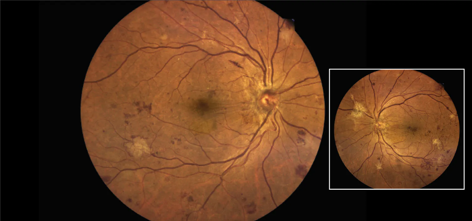
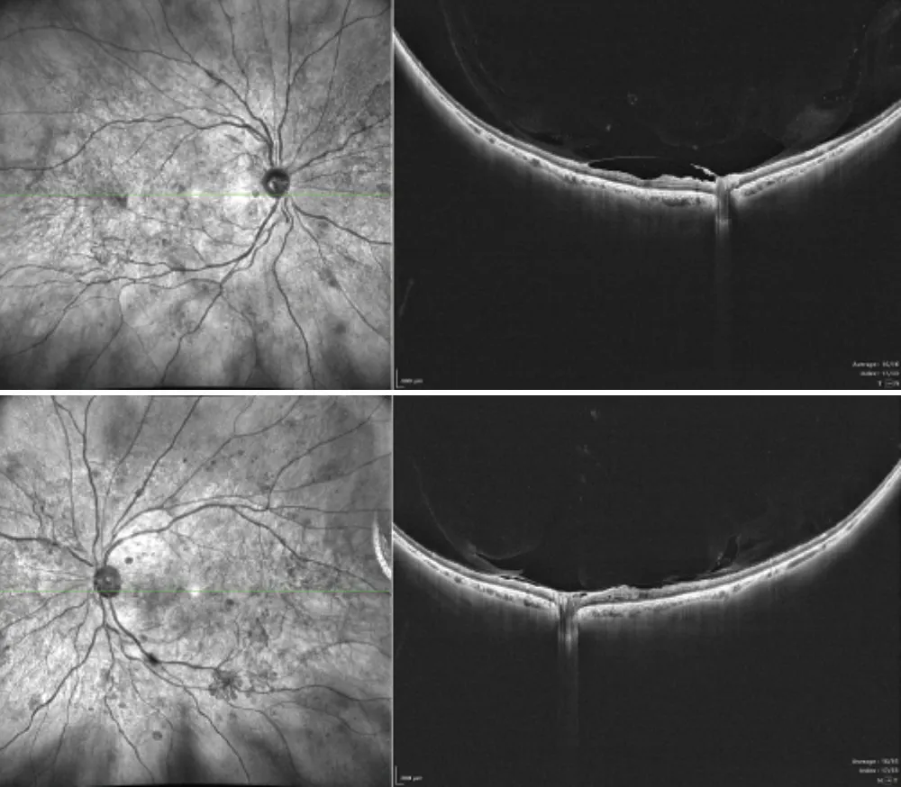
A 62-year-old man with a history of diabetes for the past 10 years presented with complaints of bilateral blurred vision for 2 months with a BCVA of 20/30 OU. Findings on color fundus photography were suggestive of proliferative diabetic retinopathy with fibrovascular proliferation around the optic disc and along the vascular arcades (Figure 1). Widefield OCT demonstrated mild alteration of the foveal contour with minimal parafoveal cystic spaces (Figure 2). The left eye also showed internal limiting membrane folds with vitreomacular adhesion. No significant macular edema was noted.
Widefield OCT angiography (OCTA) demonstrated extensive peripheral microvascular alterations consistent with a severe ischemic retinopathy (Figure 3). The most striking feature was the presence of large areas of capillary nonperfusion involving the mid- and far-peripheral retina bilaterally. These regions appeared as well-demarcated flow voids on widefield OCTA, indicating significant capillary dropout and retinal ischemia. At the junction of perfusion and nonperfusion, there were multiple abnormal vascular proliferations visualized as bright, irregular, high-flow networks. These frond-like vascular complexes exhibited disorganized branching and looping patterns, characteristic of neovascularization elsewhere.
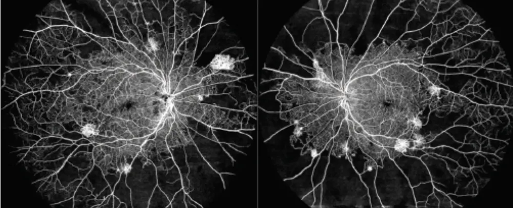
The bilateral and relatively symmetrical involvement, in conjunction with extensive peripheral capillary nonperfusion and widespread neovascularization, was highly suggestive of proliferative diabetic retinopathy. Given the extent of nonperfusion and the presence of active neovascular complexes, this patient was deemed to be at increased risk for vision-threatening complications, such as vitreous hemorrhage and tractional retinal detachment. Panretinal photocoagulation was performed in each eye with close follow-up.
MIND THE PERIPHERAL RETINA
This case demonstrates that even with relatively preserved visual acuity, advanced peripheral ischemic changes can be present, emphasizing the critical role of widefield imaging in comprehensive retinal evaluation.