




KEY TAKEAWAYS
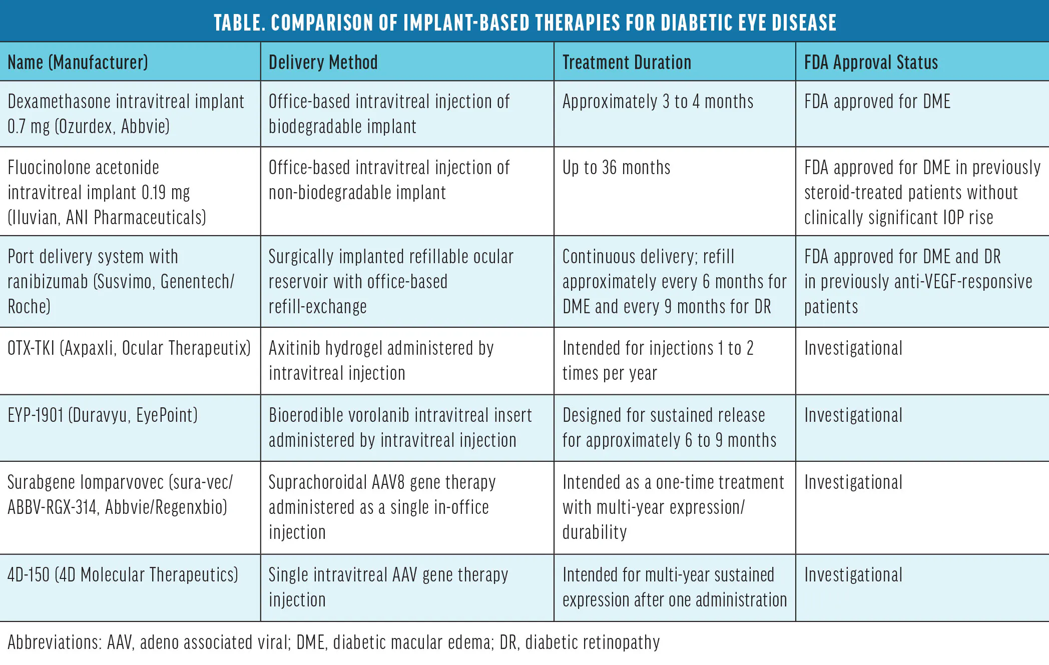
- Currently approved implants for diabetic eye disease include the dexamethasone intravitreal implant 0.7 mg (Ozurdex, Abbvie), fluocinolone acetonide intravitreal implant 0.19 mg (Iluvien, ANI Pharmaceuticals), and port delivery system with ranibizumab (Susvimo, Genentech/Roche).
- Several biodegradable polymer implants, sustained-release intraocular delivery systems, and combination antiinflammatory strategies remain under investigation.
- Surabgene lomparvovec (sura-vec/ABBV-RGX-314, Abbvie/Regenxbio) and 4D-150 (4D Molecular Therapeutics) are both in phase 2 for diabetic macular edema.
Diabetic eye disease (DED) continues to be a leading cause of vision impairment among working-age adults worldwide, and the burden of diabetic retinopathy (DR) and diabetic macular edema (DME) on patients and health care systems is expected to increase substantially. Conventional treatments—including focal/grid laser photocoagulation, intravitreal anti-VEGF agents, and corticosteroids—have significantly improved visual outcomes over the past 2 decades. However, many patients experience incomplete responses, require frequent injections, or struggle with adherence due to treatment burden or access limitations.1-3
Implantable drug-delivery platforms have emerged as an important evolution in the management of DED. By enabling sustained intraocular therapeutic exposure, implants are designed to reduce treatment frequency, improve durability of response, and better match the chronic, relapsing nature of DED. Early corticosteroid implants have now been followed by refillable anti-VEGF reservoirs and investigational gene-based and biomaterial-driven approaches, reshaping long-term treatment strategies. Here, we summarize currently approved implants, emerging technologies, and future directions in sustained therapy for DED (Table).
APPROVED IMPLANT OPTIONS
Several intravitreal implants are currently available for the treatment of DME, primarily using corticosteroid or anti-VEGF delivery to target inflammatory and vascular permeability pathways known to contribute to DME. These devices differ in biodegradability, duration of drug release, and suitability for specific patient populations.1,4
Intravitreal Corticosteroid Implants
Corticosteroids exert beneficial effects in DME by inhibiting inflammatory cytokines, stabilizing the blood-retinal barrier, and downregulating VEGF expression.1 The dexamethasone intravitreal implant 0.7 mg (Ozurdex, Abbvie) is a biodegradable polymer-based implant that delivers sustained corticosteroid exposure for approximately 3 to 4 months. Multiple randomized and real-world studies have demonstrated significant improvements in BCVA and reductions in central retinal thickness in patients with DME, including those previously treated with anti-VEGF agents.5-7
The dexamethasone implant has shown utility in pseudophakic eyes, vitrectomized eyes, and patients with inflammatory-predominant DME or suboptimal anti-VEGF response (Figure).1,6 Adverse effects such as IOP elevation and cataract progression are well characterized and generally manageable with appropriate monitoring.5-7
The fluocinolone acetonide intravitreal implant 0.19 mg (Iluvien, ANI Pharmaceuticals) represents a longer-acting, non-biodegradable corticosteroid platform approved for chronic DME insufficiently responsive to other treatments. This implant delivers low-dose fluocinolone acetonide continuously for up to 36 months. The pivotal FAME trials demonstrated sustained visual improvement and edema control over 3 years, with durable anatomic benefits and meaningful reductions in treatment burden.8
Long-term real-world studies, including the PALADIN study, have confirmed sustained efficacy and a favorable safety profile in carefully selected patients who have demonstrated steroid tolerance.9 Cataract formation and IOP elevation remain common, underscoring the importance of patient selection and pre-implant steroid challenge.8,9
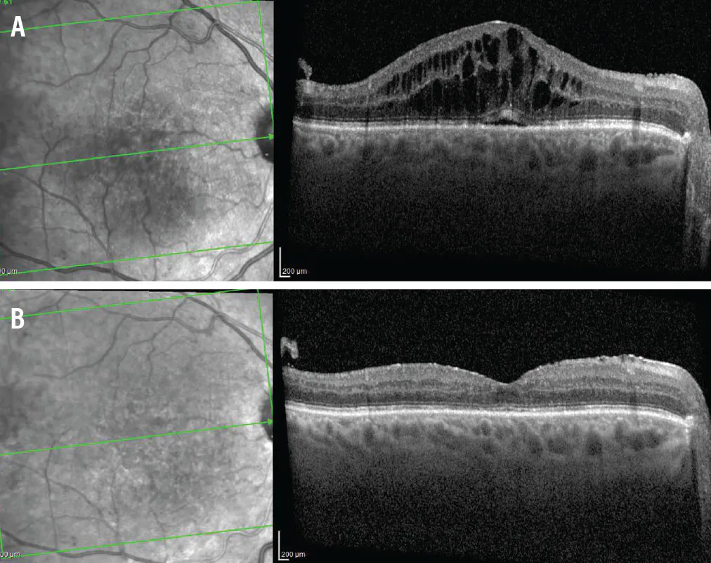
Figure. This patient with DME was not responding well to anti-VEGF therapy (A), necessitating the addition of a dexamethasone implant (B).
Intravitreal Reservoir
The port delivery system (PDS) with ranibizumab (Susvimo, Genentech/Roche) is a refillable, surgically implanted reservoir that continuously releases ranibizumab into the vitreous cavity and is periodically refilled via a minimally invasive office-based procedure.
Clinical studies of the PDS have demonstrated the feasibility of extended anti-VEGF delivery with reduced treatment frequency in retinal disease, supporting the broader concept of durable refillable intraocular biologic therapy. However, disease-specific evidence in DME is still evolving, and safety considerations such as conjunctival complications, implant-related adverse events, and endophthalmitis remain important for patient counseling and long-term monitoring.10,11
BEYOND SUSTAINED DELIVERY: CAN WE MEASURE WHAT'S LEFT?
A noninvasive method for measuring residual drug could lead to pharmacologically guided care.
By Gennady Landa, MD, and Gennady Friedman, PhD


When managing diabetic eye disease, refillable anti-VEGF reservoirs and sustained-release corticosteroid implants can reduce injection frequency and treatment burden while maintaining therapeutic exposure.1-3 Yet one clinically important question remains unanswered at follow-up visits: How much active drug is left inside the implant?
Currently, retina specialists rely on predetermined retreatment schedules and indirect evidence of declining therapeutic effect, such as recurrent fluid on OCT, worsening visual acuity, or disease progression. This reactive approach may expose some patients to recurrence before retreatment, while subjecting others to retreatment procedures or supplemental therapy despite adequate remaining drug. It can also make it difficult to distinguish drug depletion from other causes of suboptimal response.
A newly developed proprietary technology platform, developed through a collaboration between the Department of Ophthalmology at the Icahn School of Medicine at Mount Sinai and the College of Engineering and Computing at Drexel University, may offer another approach. The concept incorporates engineered magnetic nanoparticles confined within the implant matrix and uses an external reader to obtain a noninvasive signal associated with the amount of drug remaining. In principle, the measurement could be performed during a routine office visit and provide quantitative information to complement OCT, visual acuity, and clinical examination. A low residual drug measurement could favor retreatment, whereas an adequate measurement with disease activity may prompt consideration of an alternative mechanism or adjunctive therapy.
While this is a promising concept, many hurdles to clinical translation remain, including validation of measurement accuracy, signal stability over time, biocompatibility, nanoparticle confinement, compatibility with implant manufacturing, and performance across different therapeutic agents, implant materials, geometries, positions, and tissue overgrowth or fibrosis. Finally, prospective studies must show the benefits of real-time monitoring.
For retina specialists managing diabetic eye disease, the ability to measure what remains inside an implant could represent the next step toward precision dosing and more proactive disease control.
Gennady Landa, MD
Professor of Ophthalmology, Director of the Retina Service, Department of Ophthalmology, New York Eye and Ear Infirmary of Mount Sinai, Icahn School of Medicine, New York, New York
doctor.landa@gmail.com
Financial disclosure: Intellectual Property (magnetic nanoparticle-based monitoring technology)
Gennady Friedman, PhD
Professor of Electrical and Computer Engineering, College of Engineering and Computing, Drexel University, Philadelphia
Financial disclosure: Intellectual Property (magnetic nanoparticle-based monitoring technology)
AI disclosure: ChatGPT 5.6 (OpenAI) was used to assist with language editing and manuscript formatting. All content was reviewed, verified, and revised by the authors.
1. Khanani AM, Campochiaro PA, Graff JM, et al. Continuous ranibizumab via port delivery system vs monthly ranibizumab for treatment of diabetic macular edema: The Pagoda randomized clinical trial. JAMA Ophthalmol. 2025;143(4):326-335. doi.org/10.1001/jamaophthalmol.2025.0006
2. Campochiaro PA, Brown DM, Pearson A, et al; FAME Study Group. Sustained delivery fluocinolone acetonide vitreous inserts provide benefit for at least 3 years in patients with diabetic macular edema. Ophthalmology. 2012;119(10):2125-2132. doi.org/10.1016/j.ophtha.2012.04.030
3. Susvimo (ranibizumab injection) [prescribing information]. Genentech. 2025.
INVESTIGATIONAL IMPLANTS
The future of implant therapy for DED is increasingly focused on long-term disease modification and novel biologic approaches to address both the vascular and neurodegenerative aspects of DED.4,12 Hydrogel-based systems and nanotechnology-enabled platforms are being explored to improve drug stability, prolong release, and enhance tissue targeting in retinal disease.12,13,16 Additional innovation is occurring in platforms designed for controlled release, improved ocular bioavailability, and multimodal treatment delivery. As these technologies mature, cost-effectiveness analyses and real-world durability studies will be critical to define their role alongside established treatment paradigms.12,13,16
Tyrosine Kinase Inhibitors
Sustained-release tyrosine kinase inhibitor (TKI) implants are an investigational strategy for DR and DME. Unlike extracellular anti-VEGF agents, TKIs act intracellularly to inhibit VEGF receptor signaling and may also affect related angiogenic and inflammatory pathways implicated in vascular leakage and neovascularization.
OTX-TKI (Axpaxli, Ocular Therapeutix), an axitinib-containing bioresorbable hydrogel implant, is being evaluated in phase 3 for nonproliferative DR, with dosing strategies that include single or repeat administration at 24 weeks.
EYP-1901 (Duravyu, EyePoint), a bioerodible intravitreal insert containing vorolanib, is under investigation in two phase 3 trials for the treatment of DME. These platforms are intended to reduce injection burden while maintaining durable suppression of VEGF-driven permeability and disease progression.19-21
For more on TKIs, see The Potential of Tyrosine Kinase Inhibitors.
Gene Therapy
Gene-based delivery systems represent a rapidly evolving investigational category. These treatments typically use viral vectors to promote sustained intraocular expression of therapeutic proteins after a single administration. Contemporary reviews highlight the promise of ocular gene therapy while also emphasizing manufacturing, durability, and inflammatory safety challenges that must be addressed before broader clinical adoption.14,15
In DED, most clinical gene therapy strategies have focused on sustained intraocular suppression of VEGF signaling. Adeno-associated viral (AAV) vectors are the predominant platform used, as they can drive prolonged retinal expression after a single administration. These gene therapies largely target VEGF-A directly through viral-mediated expression of anti-VEGF proteins; however, newer therapies are also investigating inhibition of VEGF-C, placental growth factor, and other mediators of vascular permeability and inflammation. Routes of delivery vary by platform and include intravitreal, suprachoroidal, and subretinal administration, each with tradeoffs in transduction efficiency, procedural complexity, and inflammatory risk.14-16
Surabgene lomparvovec (sura-vec/ABBV-RGX-314, Abbvie/Regenxbio) is an AAV8-based therapy encoding an anti-VEGF antibody fragment administered suprachoroidally for DME.17 Early data from the phase 2 ALTITUDE trial have been encouraging.17
Early data from the phase 2 SPECTRA trail of 4D-150 (4D Molecular Therapeutics) is also promising.18 This intravitreal gene therapy is designed to potentially provide multi-year expression of an aflibercept-like anti-VEGF moiety and an inhibitory RNA molecule that blocks intracellular expression of VEGF-C.
ADVANTAGES, LIMITATIONS, AND PATIENT SELECTION
Implant-based therapies offer several advantages over intravitreal injections, including reduced treatment burden, improved adherence potential, and more consistent pharmacokinetics.3,12 These benefits are particularly relevant for patients with limited access to care, poor injection tolerance, or chronic/persistent edema requiring frequent retreatment.
However, implants are not without risk. Surgical implantation, device-specific complications, steroid-related adverse events, and higher upfront costs must all be weighed against expected benefits.1,4 Thoughtful patient selection remains paramount: Corticosteroid implants may be best suited for pseudophakic patients, those with inflammatory DME phenotypes, or eyes refractory to anti-VEGF therapy while anti-VEGF implants may offer broader applicability across different disease stages.6,8,10
UPDATING THE PARADIGM
Implantable drug delivery systems represent a transformative advance in the management of DR and DME. With multiple approved corticosteroid implants and expanding anti-VEGF and investigational platforms, retina specialists now have unprecedented opportunities to individualize therapy and reduce treatment burden. Careful patient selection, vigilant monitoring, and continued evaluation of long-term outcomes remain essential as emerging technologies further reshape the therapeutic landscape of DED.3,4,6
AI disclosure: Microsoft Copilot (version 148.0.3967.70) was used to assist with drafting and editing of the article. The authors reviewed the entire contents of this article and confirmed its accuracy.
Disclosure of off-label/investigational use: OTX-TKI, EYP-1901, surabgene lomparvovec, and 4D-150 are investigational therapies that are not currently FDA approved.
1. Schmidt-Erfurth U, Garcia-Arumi J, Bandello F, et al. Guidelines for the management of diabetic macular edema by the European Society of Retina Specialists (EURETINA). Ophthalmologica. 2017;237(4):185-222. doi.org/10.1159/000458539
2. Campochiaro PA, Brown DM, Pearson A, et al. Sustained delivery fluocinolone acetonide vitreous inserts provide benefit for at least 3 years in patients with diabetic macular edema. Ophthalmology. 2012;119(10):2125-2132. doi.org/10.1016/j.ophtha.2012.04.030
3. Campochiaro PA, Hafiz G, Shah SM, et al. Sustained ocular delivery of fluocinolone acetonide by an intravitreal insert. Ophthalmology. 2010;117(7):1393-1399.e3. doi.org/10.1016/j.ophtha.2009.11.024
4. Syed YY. Fluocinolone acetonide intravitreal implant 0.19 mg (Iluvien): A review in diabetic macular edema. Drugs. 2017;77(5):575-583. doi.org/10.1007/s40265-017-0722-4
5. Busch C, Fraser-Bell S, Iglicki M, et al. Real-world outcomes of non-responding diabetic macular edema treated with continued anti-VEGF therapy versus early switch to dexamethasone implant: 2-year results. Acta Diabetologica. 2019;56(12):1341-1350. doi.org/10.1007/s00592-019-01416-4
6. Bonfiglio V, Reibaldi M, Pizzo A, et al. Dexamethasone for unresponsive diabetic macular oedema: optical coherence tomography biomakers. Acta Ophthalmologica. 2019;97(4): e540-e544. doi.org/10.1111/aos.13935
7. Guigou S, Pommier S, Meyer F, et al. Efficacy and safety of intravitreal dexamethasone implant in patients with diabetic macular edema. Ophthalmologica. 2015;233(3-4):169-175. doi.org/10.1159/000381356
8. Campochiaro PA, Brown DM, Pearson A, et al. Long-term benefit of sustained-delivery fluocinolone acetonide vitreous inserts for diabetic macular edema. Ophthalmology. 2011;118(4):626-635.e2. doi.org/10.1016/j.ophtha.2010.12.028
9. Singer MA, Sheth V, Mansour SE, et al. Three-year safety and efficacy of the 0.19-mg fluocinolone acetonide intravitreal implant for diabetic macular edema: The PALADIN study. Ophthalmology. 2022;129(6):605-613. doi.org/10.1016/j.ophtha.2022.01.015
10. Holekamp NM, Campochiaro PA, Chang MA, et al. Archway randomized phase 3 trial of the port delivery system with ranibizumab for neovascular age-related macular degeneration. Ophthalmology. 2022;129(3):295-307. doi.org/10.1016/j.ophtha.2021.09.016
11. Campochiaro P, Marcus D, Awh C, et al. The port delivery system with ranibizumab for neovascular age-related macular degeneration. Ophthalmology. 2019; 126(8):1141-1154. doi.org/10.1016/j.ophtha.2019.03.036
12. Urtti A. Challenges and obstacles of ocular pharmacokinetics and drug delivery. Adv Drug Deliv Rev. 2006;58(11):1131-1135. doi.org/10.1016/j.addr.2006.07.027
13. Yasukawa T, Ogura Y, Sakurai E, et al. Intraocular sustained drug delivery using implantable polymeric devices. Adv Drug Deliv Rev. 2005;57(14):2033-2046. doi.org/10.1016/j.addr.2005.09.005
14. Rodrigues GA, Shalaev E, Karami TK, et al. Pharmaceutical development of AAV-based gene therapy products for the eye. Pharm Res. 2019;36(2):29. doi.org/10.1007/s11095-018-2554-7
15. Trapani I, Auricchio A. Seeing the light after 25 years of retinal gene therapy. Trends Mol Med. 2018;24(8):669-681. doi.org/10.1016/j.molmed.2018.06.006
16. Rowe LW, Ciulla TA. Gene therapy for non-hereditary retinal disease: age-related macular degeneration, diabetic retinopathy, and beyond. Genes. 2024;15(6):720. doi.org/10.3390/genes15060720
17. RGX-314 gene therapy administered in the suprachoroidal space for participants with diabetic retinopathy (DR) with and without center involved-diabetic macular edema (CI-DME) (ALTITUDE). Updated September 17, 2025. clinicaltrials.gov/study/NCT04567550
18. 4D-150 in patients with diabetic macular edema (SPECTRA). Updated September 22, 2025. clinicaltrials.gov/study/NCT05930561
19. A study to evaluate the efficacy and safety of OTX-TKI (axitinib implant) in participants with non-proliferative diabetic retinopathy (HELIOS-3). ClinicalTrials.gov. Accessed July 8, 2026. clinicaltrials.gov/study/NCT07235085
20. COMO: a phase 3 randomized, double-masked study comparing the efficacy of EYP-1901 against aflibercept in DME (DME). ClinicalTrials.gov. Accessed July 8, 2026. clinicaltrials.gov/study/NCT07449936
21. CAPRI: a phase 3 randomized, double-masked study comparing the efficacy of EYP-1901 against aflibercept in DME (DME). ClinicalTrials.gov. Accessed July 8, 2026. clinicaltrials.gov/study/NCT07449923


