




A 69-year-old man presented to the retina clinic for consultation of a chronic, refractory macular hole (MH). He had a history of nonproliferative diabetic retinopathy and a rhegmatogenous retinal detachment repaired more than 10 years ago with a scleral buckle, pars plana vitrectomy, and silicone oil tamponade. Despite successful reattachment of the retina, he developed a chronic MH. He later underwent silicone oil removal, and the chronic MH enlarged. His VA was 20/400 in the affected eye.
OCT imaging showed a full-thickness MH, moth-eaten appearance of the MH edges with loss of the ellipsoid and interdigitation zones (indicative of chronicity), cystoid spaces, inner retinal dimpling (from prior membrane peeling), and patchy attenuation of the retinal nerve fiber layer (Figure 1A).
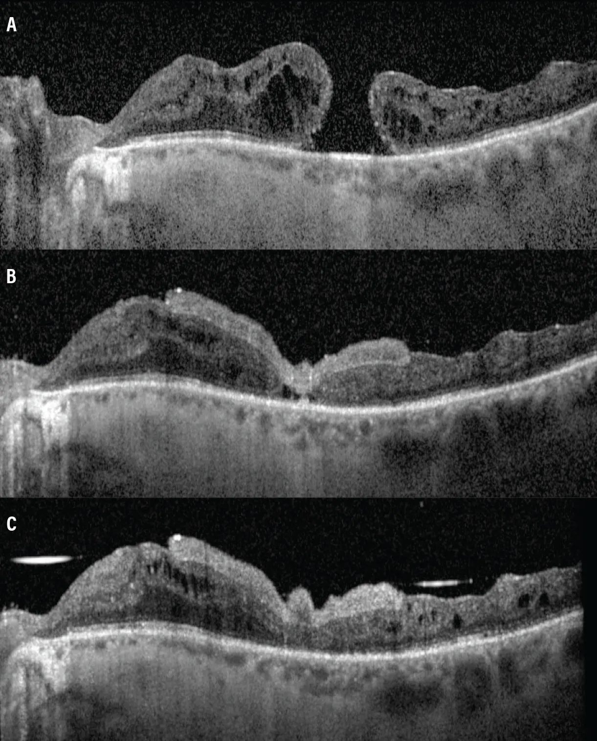
Figure 1. Preoperative OCT imaging shows the MH at presentation (A). Postoperative month 1 shows preretinal AMT without full apposition of the MH edges (B). OCT imaging at postoperative month 2 shows improvement and closure of the MH (C).
The patient underwent 25-gauge pars plana vitrectomy, brilliant blue-assisted epiretinal membrane and internal limiting membrane peeling, MH massage, and a 4 mm preretinal amniotic membrane transplant (AMT) under perfluorocarbon liquid (PFCL), which was then directly exchanged for 1,000 cS silicone oil (Figure 2).
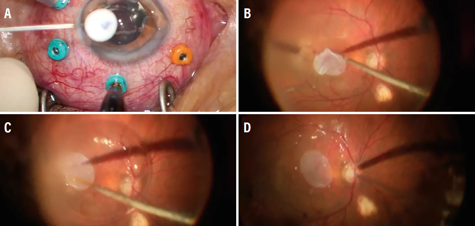
Figure 2. Intraoperative imaging shows a cryopreserved human AMT prepared using a 4 mm circular punch with a gentian violet “F” stamp marking the epithelial side, use of chandelier illumination for bimanual manipulation, and use of 23-gauge cannula for easier insertion of the AMT (A). We unfurled the AMT on the macular surface using a bimanual technique with a Finesse flex loop (Alcon) and internal limiting membrane forceps (B). We then used PFCL to stabilize the AMT (C), followed by direct PFCL-silicone oil exchange (D).
OCT imaging at postoperative months 1 and 2 demonstrated MH closure with a well-positioned preretinal AMT (Figure 1B and C). The patient’s VA improved to 20/200 in the affected eye by postoperative month 7, and he reported a subjective improvement in vision and reduction in central scotoma size. The silicone oil and preretinal amniotic membrane were left in place.
AMT TO THE RESCUE
AMTs may be effective for refractory MHs (closure rates 85-100%), myopic MH associated with posterior staphyloma (86-92%), and complex retinal detachments with proliferative vitreoretinopathy (76-92%).1 Visual improvement was reported in 81% of cases in a review of 603 eyes; however, the extent of improvement is highly variable.2 The most common complication is graft dislocation (3.6%).2 Further large-scale prospective studies are needed to better compare AMT with established techniques for MH repair.
1. Carlà MM, Giannuzzi F, Hu L, et al. The human amniotic membrane in vitreoretinal surgery: Applications, outcomes and limitations. Surv Ophthalmol. 2026;71(1):14-24. doi.org/10.1016/j.survophthal.2025.08.004
2. Solish D, Gao A, Cheffi N, Mandelcorn E, Felfeli T. Human amniotic membrane (hAM) in macular hole repair: a scoping review [published online ahead of print May 6, 2026]. Retina. doi.org/10.1097/IAE.0000000000004877


