



Multimodal imaging is central to managing patients with geographic atrophy (GA) because each method used in the clinic contributes distinct information for diagnosis, risk stratification, treatment discussions, longitudinal monitoring, and education. Together, the various modalities provide a more complete understanding of GA than any single imaging method alone.
 What Each Imaging Modality Adds
Each imaging modality offers a different perspective on GA and contributes unique information to the treatment conversation.
- Structural OCT serves as a foundational imaging modality for identifying early disease changes and monitoring progression over time. OCT helps to identify features such as nascent GA, incomplete retinal pigment epithelium and outer retina atrophy (iRORA), hyperreflective wedge-shaped bands, changes in the outer plexiform layer (OPL) and inner nuclear layer, and foveal involvement.
- Fundus autofluorescence (FAF) helps visualize lesion characteristics and supports both diagnosis and patient education. This modality is often used to distinguish AMD-related GA from lookalikes and help patients understand disease progression visually. For example, FAF may reveal lesion size and configuration, multifocal lesions, perilesional hyperautofluorescence, and features suggestive of inherited retinal disease mimickers of GA.
- OCT angiography provides noninvasive vascular assessment and adds vascular information that complements structural imaging. This imaging is used to evaluate for choroidal neovascularization, which can occur with increased frequency in patients with GA, and assess vascular changes without fluorescein dye.
- Color fundus photography remains useful for documenting visible pathology and facilitating patient communication. However, it is limited in its ability to detect early GA, identify subtle lesions, visualize non-exudative neovascularization, and detect high-risk findings such as reticular pseudodrusen/subretinal drusenoid deposits. Therefore, fundus photography alone is not sufficient for a comprehensive GA evaluation.
 Imaging Frequency and Monitoring Approach
Imaging frequency varies according to disease severity, progression risk, and treatment status. In my experience, I collect images at the following intervals and disease progression points:
- OCT and FAF every 6 months for patients being followed with GA without treatment
- Imaging every 3 to 4 months for higher-risk patients or those considering therapy
- OCT at each injection visit during complement inhibitor treatment
- FAF approximately every 6 months during treatment during a dilated exam to facilitate improved image capture
These imaging intervals provide additional data points over time, which can help monitor disease progression, identify concerning changes, and support ongoing treatment discussions (Figure).
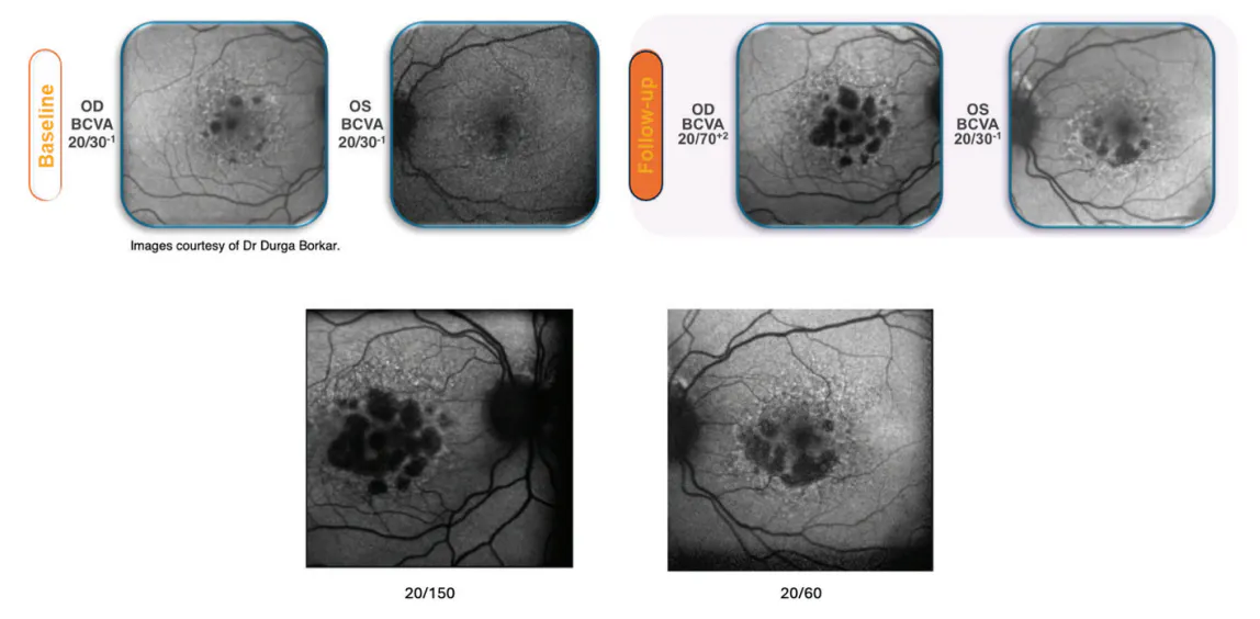
Figure. FAF of a 74-year-old woman with progressive extrafoveal multifocal GA OD > OS. FAF shows continued progression over time prior to initiating complement inhibition when available.
 How Biomarkers Inform Treatment Discussions
When collecting patient images, I look out for several biomarkers that can suggest increased risk for GA progression. These include reticular pseudodrusen, hyperreflective foci, perilesional hyperautofluorescence, multifocal and extrafoveal lesions, and larger baseline lesion size. Because biomarker identification helps stratify progression risk and guide follow-up intensity, these findings can help prompt treatment conversations. It is important to remember that treatment decisions extend beyond imaging findings alone and include considering whether GA is the major cause of vision impairment, the presence of other ocular comorbidities such as advanced glaucoma, and patient willingness and ability to sustain ongoing injection therapy.
 Emerging Role of AI in GA Imaging
In many areas of medicine, specifically ophthalmology, AI-based imaging analysis is increasingly moving toward clinical implementation. Currently, potential AI applications in practice include automated lesion measurement, monitoring lesion growth, segmentation analysis, hypertransmission mapping, and quantification of retinal layer atrophy. In my opinion, however, the science still seems to be ahead of the implementation in routine clinical care. As AI capabilities continue to evolve, certain barriers to workflow integration remain. These include device-specific variability, differences in OCT image quality, limited interoperability, and incomplete EHR integration. Addressing these barriers may be important for integrating AI-based imaging analysis into routine GA care.
Closing Takeaway
Multimodal imaging forms the basis of modern GA management. Structural, autofluorescence, vascular, and photographic imaging each contribute important, complementary information that supports a multitude of aspects including diagnosis, prognosis, and treatment planning.


_1784132761.jpg?auto=compress,format&w=70)


