



KEY TAKEAWAYS
- The 54th annual Aspen Retinal Detachment Society (ARDS) meeting was held February 28 – March 4, in Snowmass, Colorado.
- The event included three panel discussions on surgical retina, the drug pipeline, and complex cases.
- The 55th annual ARDS meeting is scheduled for February 27 – March 3, 2027, in Snowmass, Colorado.
The 54th annual Aspen Retinal Detachment Society (ARDS) meeting, held February 28 – March 4, in Snowmass, Colorado, brought together leaders in vitreoretinal surgery, medical retina, ocular oncology, and uveitis for a series of lively panel discussions. As always, these sessions generated thoughtful debate, practical clinical pearls, and valuable perspectives. Here, I summarize the meeting’s three engaging panels, which focused on surgical decision making and emerging pharmacologic therapies.
PANEL 1: ADVANCES IN SURGICAL RETINA
The first panel, moderated by Timothy G. Murray, MD, MBA, opened with a case of a patient with a visually significant cataract and a malignant choroidal melanoma. The panelists—including Carl C. Awh, MD; Jacque L. Duncan, MD; Allen C. Ho, MD; and Aleksandra V. Rachitskaya, MD—discussed combined versus staged surgery (Figure 1). They agreed that the approach depends on the pathology, surgeon expertise, and the availability of local cataract surgeons. Dr. Ho noted that if the tumor is growing, it might be preferable to address the malignancy first. Dr. Awh suggested that patients with epiretinal membrane and vitreomacular traction often gain enough improvement from cataract surgery that they do not need macular surgery. The panelists also briefly touched on the use of dyes for macular surgery; most use indocyanine green (ICG) dye and have not had cases of ICG-related phototoxicity.
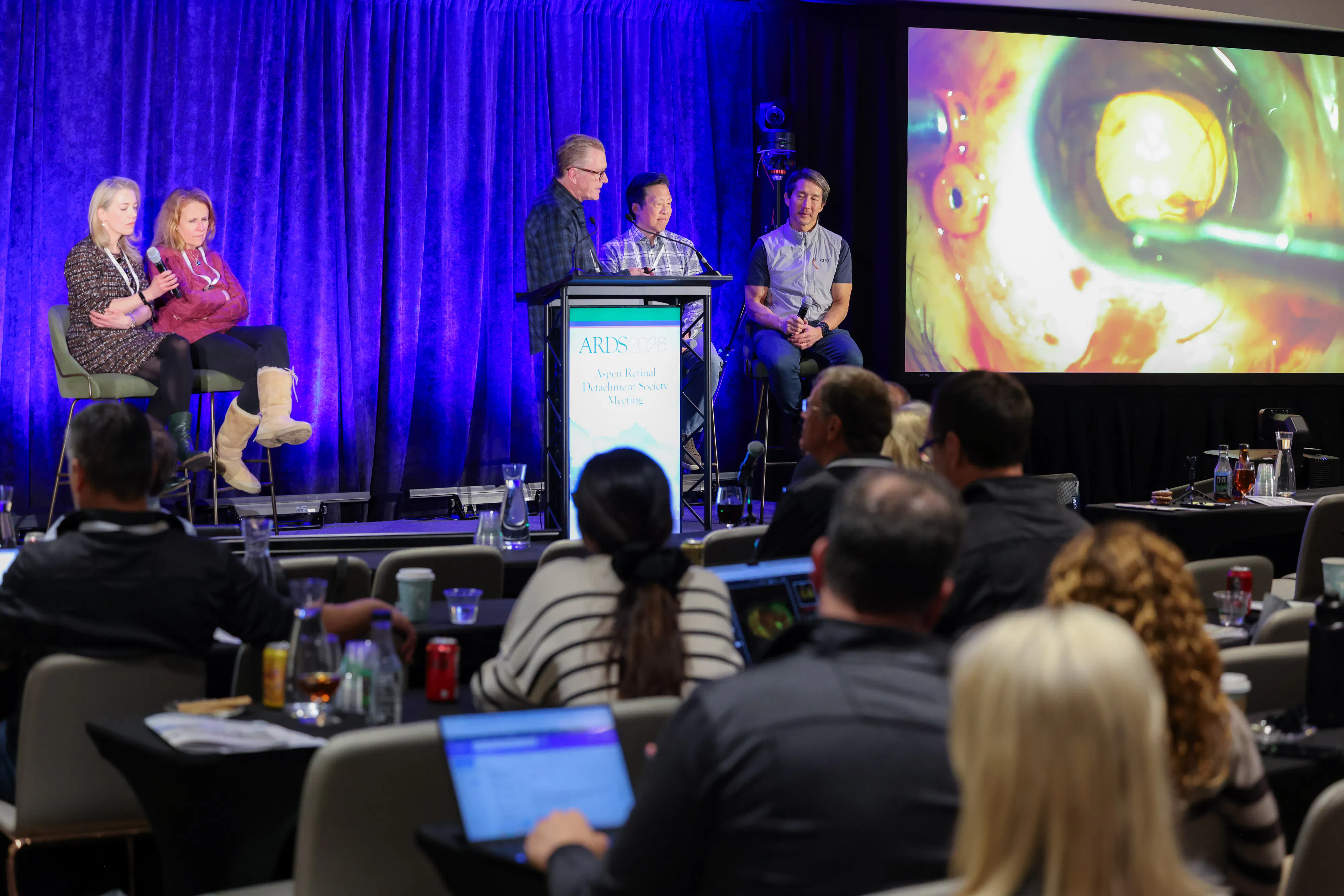
Figure 1. The first panel showcased the expertise of Drs. Rachitskaya, Duncan, Murray, Awh, and Ho. Image Courtesy of Kevin Caldwell Photography.
The second case of a dislocated IOL generated discussion about secondary lens options. The panelists stressed the importance of understanding lens type, capsular support, and patient-specific factors before selecting a surgical approach. The group acknowledged the many surgical techniques available, and surgeons should use the technique that gives them the best surgical outcome based on their own experience and training. Evolving secondary IOL technologies were briefly discussed, including an investigational prosthetic capsular support system.
A case of epiretinal membrane highlighted the variable use of dye, routine internal limiting membrane peeling, and combined phacoemulsification. Finally, Dr. Murray shared a case of iris melanoma with ciliary body extension, in which he completed cataract extraction, IOL implantation, and intraoperative endolaser to the tumor, emphasizing the importance of lifting the hyaloid in these cases.
PANEL 2: DRUG TREATMENTS WE USE AND THE PIPELINE
The second panel, moderated by Donald J. D’Amico, MD, focused on one of retina’s most persistent challenges: extending treatment durability while maintaining visual outcomes. The panel included Dr. Duncan; Tarek S. Hassan, MD; Tara A. McCannel, MD, PhD; Lucia Sobrin, MD, MPH; and Robert L. Avery, MD (Figure 2). The discussion opened with the port delivery system (PDS) with ranibizumab (Susvimo, Genentech/Roche), which has demonstrated excellent visual acuity preservation through sustained anti-VEGF delivery. Despite these results, the panelists agreed that widespread adoption has been limited by practical considerations, including surgical implantation, reimbursement challenges, and the growing availability of longer-acting injectable agents. Dr. D’Amico pointed out that our perceptions of the PDS technology might have been different had every-6-month surgical implants preceded monthly intravitreal injections. The panelists then highlighted emerging therapies designed to extend intraocular drug durability through binding to vitreous components, including TH103, an anti-VEGF biologic under development by Kalaris Therapeutics.
Drawing parallels to sustained delivery in other spaces, Dr. Sobrin shared insights from uveitis management. While longer-acting fluocinolone injectables are available, patients often still require a dexamethasone implant (Ozurdex, Abbvie) every 2 to 3 months because of its superior potency. Consequently, even longer-acting alternatives do not always reduce treatment frequency. The panelists also discussed intravitreal steroids as adjunctive therapy for diabetic macular edema, cystoid macular edema, and wet AMD, particularly in patients with incomplete responses to anti-VEGF treatment. Insurance-mandated step therapy remains a major determinant of treatment selection.
The panelists also discussed the distinction between anatomic and functional outcomes. Although newer agents may achieve greater retinal drying and longer durability, visual acuity gains have remained similar across therapies. Treatment burden, disease control, and durability should also be considered important measures of efficacy, not just visual acuity.
The session concluded with a look toward future therapeutic targets beyond VEGF inhibition, including Wnt signaling and TIE2 activation pathways, as well as the growing role of home OCT monitoring. The panelists envisioned a future in which more durable therapies are paired with remote monitoring technologies, allowing treatment to become more personalized and less burdensome for patients.
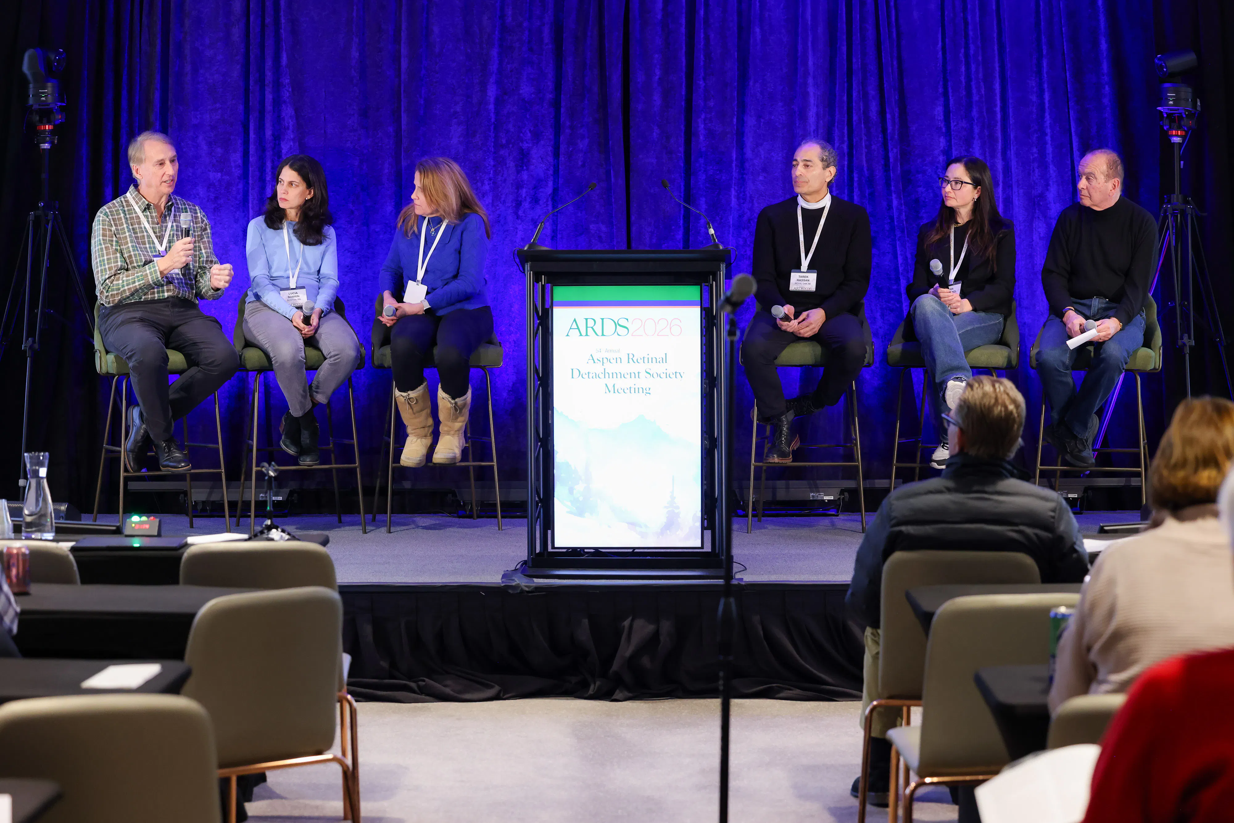
Figure 2. The second panel focused on pharmacologic therapy, with Drs. Avery, Sobrin, Duncan, Hassan, McCannel, and D’Amico. Image courtesy of Kevin Caldwell Photography.
PANEL 3: SURGICAL RETINA: TRAUMA, UVEITIS, AND VISUALIZATION
The final panel, moderated by R.V. Paul Chan, MD, MBA, MSc, and including Drs. Avery, McCannel, Sobrin, and David R. Chow, MD, explored the complexities of surgical decision making through a series of challenging cases. The first case involved a zone 1 open globe injury with an intraocular foreign body and retinal detachment (RD). Management strategies varied among the panelists: Some advocated delaying RD repair to allow the cornea to heal, while others favored immediate repair despite the potential need for a combined penetrating keratoplasty to achieve adequate visualization. Panelists also debated whether or not to add a scleral buckle. The discussion underscored the importance of flexibility in ocular trauma surgery and the role of ultrasound in the preoperative assessment.
The panel also discussed an unusual case of pediatric exudative RD that was ultimately diagnosed as uveal melanoma. The discussion served as a reminder that ocular tumors should remain in the differential even in atypical age groups and highlighted the value of multimodal imaging and subspecialty consultation when presentations do not fit classic patterns.
The panel emphasized a stepwise approach to managing uveitis and intraocular lymphoma cases. Using cases of epiretinal membranes with cystoid macular edema in uveitis, Dr. Sobrin explained that macular edema should first be treated medically, with surgery reserved for persistent traction after inflammation is controlled. In cases suspicious for primary vitreoretinal lymphoma, panelists agreed that clinicians should obtain a brain MRI before biopsy, collaborate with neuro-oncology early, and recognize the limited yield of lumbar puncture. Debate centered on diagnostic vitrectomy alone versus in combination with subretinal biopsy to maximize diagnostic yield and avoid repeat surgery.
The session concluded with a debate surrounding pneumatic retinopexy and the increasing effect of OR access on RD management. Experiences from Canada highlighted how resource limitations have driven broader use of pneumatic and temporizing “bubble-and-book” strategies, prompting discussion about how health system constraints increasingly influence surgical decision making in retina practice.
DON’T MISS THE DISCUSSION NEXT YEAR
The open discussion between panelists and attendees regarding the challenges facing retina specialists remains a hallmark of the ARDS meeting. Mark your calendars for next year’s meeting, which will deliver another set of outstanding panels.
SAVE THE DATE
55th Annual Aspen Retinal Detachment Society
February 27 – March 3, 2027
Snowmass Village, Colorado


