


KEY TAKEAWAYS
- Cancer-associated retinopathy (CAR) is a rare paraneoplastic autoimmune retinopathy characterized by progressive vision loss, for which there is no standard treatment approach.
- The authors describe a case of CAR in a patient with thymic carcinoma who was treated with an aggressive, multimodal approach including high-dose systemic corticosteroids, intravenous immunoglobulin, sub-Tenon corticosteroids, plasmapheresis, rituximab, intravitreal dexamethasone implant (Ozurdex, Abbvie), and systemic chemotherapy.
- Despite a poor prognosis, the patient experienced an improvement in functional vision, highlighting both a potential dissociation between structural damage and functional capacity and the importance of aggressive treatment.
Cancer-associated retinopathy (CAR) is a rare paraneoplastic autoimmune retinopathy characterized by progressive vision loss. It develops from the cross-reactivity of tumor antigens and retinal proteins, leading to photoreceptor and bipolar cell damage.1-3 First described in association with small-cell lung cancer, CAR has since been reported with gynecologic, hematologic, breast, prostate, and hepatocellular cancers.4 The diagnosis remains challenging, however, as many patients exhibit minimal fundoscopic abnormalities; thus, the combination of imaging and antibody testing can aid in accurate diagnosis. There is no standardized treatment strategy for CAR due to its rarity and heterogeneous presentation.
Here, we present a case of CAR in thymic carcinoma treated with a multimodal approach, highlighting the value of aggressive intervention despite profound vision loss.
CASE REPORT
A 54-year-old man presented to an outside facility with rapidly progressive, painless, bilateral vision loss over 1 week. His VA was hand motion OU and IOP was 8 mm Hg OU with no relative afferent pupillary defect. The anterior segment examination was unremarkable, while the fundus examination revealed attenuated, sclerotic ghost vessels and temporal optic disc pallor in each eye. Neuroimaging revealed an acute posterior left temporal white matter infarct, which did not adequately explain his vision loss. Testing for syphilis, tuberculosis, Lyme disease, and Bartonella were each negative. Angiotensin-converting enzyme, lysozyme, homocysteine, methylmalonic acid, vitamin B9 and B12, lupus anticoagulant, beta-2-glycoprotein, and cardiolipin were also unremarkable.
The patient was treated empirically with high-dose intravenous methylprednisolone (IVMP) 1 g daily for 5 days and underwent paraneoplastic panel testing as well as CT imaging of the chest, abdomen, and pelvis. Imaging revealed a large anterior mediastinal mass measuring 8.2 x 8.5 x 8 cm with areas of necrosis adjacent to enlarged lymph nodes; biopsy confirmed thymic carcinoma. The patient completed 5 days of IVMP with subjective improvement of vision but was lost to follow-up due to insurance issues.
He presented to our clinic 2 months later with decreased VA to light perception OU. Anterior segment and fundus examination remained unchanged (Figure 1). OCT showed significant ellipsoid zone (EZ) loss, severe photoreceptor and outer retinal atrophy, and diffuse retinal thinning in each eye (Figure 2). Fundus autofluorescence showed punctate areas of hypoautofluorescence in the macula and patchy hyperautofluorescent changes along the retinal vessels in each eye. Fluorescein angiography showed late punctate macular staining and focal filling defects surrounding vessels in each eye (Figure 3).
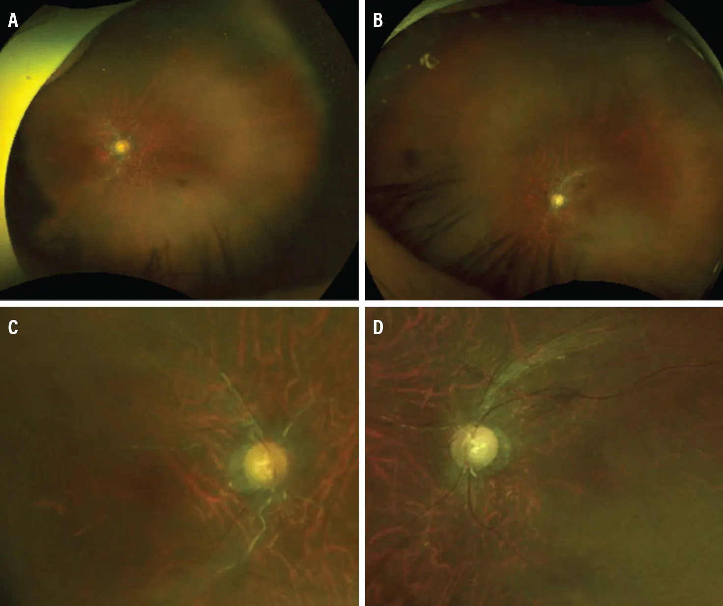
Figure 1. Fundus photography of the right (A) and left (B) eye revealed attenuated, sclerotic ghost vessels. The optic disc of the right (C) and left (D) eye demonstrated temporal pallor.
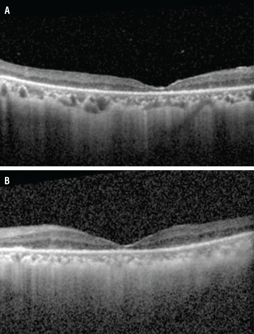
Figure 2. OCT of the right (A) and left (B) eye demonstrated significant bilateral EZ loss, severe photoreceptor and outer retinal atrophy, and diffuse retinal thinning.
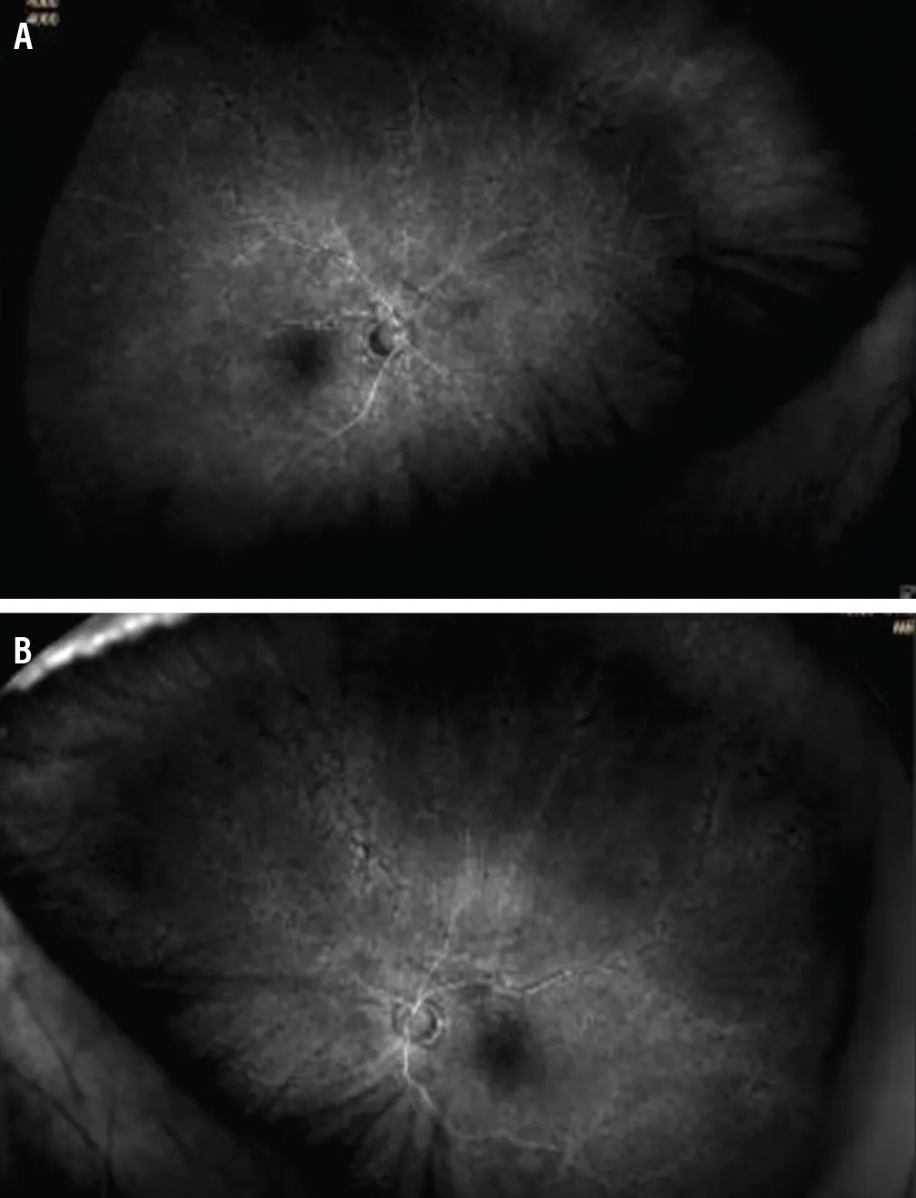
Figure 3. Fluorescein angiography of the right (A) and left (B) eye revealed late phase macular staining and focal filling defects in the superior and inferior arcades.
Given a strong suspicion of CAR and the decline in vision, a CAR panel was obtained, which was positive for recoverin, Rab6, aldolase, and enolase. He was reinitiated on IVMP 1 g daily for 5 days, followed by intravenous immunoglobulin (IVIG) 400 mg/kg/day for 5 days and sub-Tenon triamcinolone injection 40 mg with no changes in vision. Nine weeks after vision loss (10 days after presentation), systemic chemotherapy with seven cycles of paclitaxel and carboplatin was initiated, as the tumor was deemed unresectable due to proximity and possible invasion into the right atrium. Four months after vision loss, his VA remained light perception OU. A repeat sub-Tenon triamcinolone 40 mg injection was administered, followed by three sessions of plasmapheresis and four cycles of rituximab.
Five months after vision loss, the patient’s VA improved to hand motion OU. An intravitreal dexamethasone implant 0.7 mg (Ozurdex, Abbvie) was placed into each eye. Eight months after vision loss, he continued to report subjective improvement; his VA was counting fingers at 7 ft eccentrically. Repeat intravitreal dexamethasone implant was placed into each eye, and he was scheduled for external beam radiation therapy as further cancer management.
HOW LATE IS TOO LATE FOR MEANINGFUL VISUAL RECOVERY?
To the best of our knowledge, this is the first reported case of thymic carcinoma with CAR. Among the relevant autoantibodies, the anti-recoverin antibody, which was positive in this patient, is most commonly implicated in CAR and results in the most severe manifestation of the disease, as both rods and cones are targeted.5 The association with thymic carcinoma in this case broadens the spectrum of malignancies linked to CAR.
This case is also notable for the profound functional and physiologic damage to the patient’s vision at the time treatment was initiated. Early treatment is considered critical in autoimmune retinopathies, given the potential irreversible damage caused by photoreceptor apoptosis. However, our methodology of treating CAR so far is largely based on case reports, as it is difficult to conduct large-scale studies with such a rare entity. Moreover, treatment of the cancer itself does not necessarily improve the vision loss associated with CAR.6-11 The patient in this case had extensive EZ loss and outer retinal atrophy on OCT, considered to be poor prognostic indicators reflecting significant irreversible photoreceptor damage,3 and experienced a 2-month delay between initial vision loss and therapy. Nevertheless, he received high-dose systemic corticosteroids, IVIG, sub-Tenon corticosteroids, plasmapheresis, rituximab, intravitreal dexamethasone implants, and systemic chemotherapy. All these treatments have been used in case reports of CAR, but to our knowledge, no reported cases describe the use of nearly all available therapeutic interventions in a single patient.6-11 Despite the absence of a standardized treatment algorithm for CAR, this aggressive, multimodal approach was associated with functional visual improvement in this case.
It is worth noting that patients such as ours tend to experience limited, if any, meaningful recovery; nevertheless, the improvement in VA from light perception to counting fingers at 7 ft highlights a potential dissociation between structural damage and functional capacity. It is possible that OCT findings alone may not fully predict functional recovery even when imaging appears severe, suggesting autoimmune retinopathies may exhibit partial reversibility under aggressive immunomodulation and supporting the potential benefits of offering aggressive therapy, even in advanced cases with delayed presentation.
PEDAL TO THE METAL
In most cases of CAR, vision loss occurs after a malignancy diagnosis. However, in a small subset of patients, ocular findings are the first manifestation. Therefore, prompt evaluation for occult malignancy should be considered in similar cases with rapidly progressive bilateral vision loss associated with EZ and outer retinal atrophy and minimal to no intraocular inflammation.
Due to the rarity and heterogeneity in presentation of CAR, there is currently no standardized treatment strategy. In our case, even with profound vision loss and poor prognostic indicators on multimodal imaging, an aggressive treatment approach with intraocular steroids and immunomodulatory therapy lead to meaningful functional recovery.
1. Naramala S, Ahmad J, Adapa S, Gavini F, Konala VM. Case series of cancer-associated retinopathy (CAR). Cureus. 2019;11(6):e4872. doi.org/10.7759/cureus.4872
2. Singh D, Tripathy K. Cancer-associated retinopathy. February 27, 2024. In: StatPearls [Internet]. Treasure Island (FL): StatPearls Publishing; 2025.
3. Finn AP, Thomas AS, Stinnett SS, Keenan RT, Grewal DS, Jaffe GJ. The role of cystoid macular edema as a marker in the progression of non-paraneoplastic autoimmune retinopathy. Graefes Arch Clin Exp Ophthalmol. 2018;256(10):1867-1873. doi.org/10.1007/s00417-018-4084-8
4. Fang HS, Yang CS, Cheng CK, Wang YS. Cancer-associated retinopathy as an initial presentation of gynecologic small-cell carcinoma. Taiwan J Ophthalmol. 2024;15(3):494-498. doi.org/10.4103/tjo.TJO-D-23-00100
5. Weleber RG, Watzke RC, Shults WT, et al. Clinical and electrophysiologic characterization of paraneoplastic and autoimmune retinopathies associated with antienolase antibodies. Am J Ophthalmol. 2005;139(5):780-794. doi.org/10.1016/j.ajo.2004.12.104
6. Mudri J, Phagura RS, Lam WS, Wu XN. Intravitreal dexamethasone implant use as first-line therapy for cancer-associated retinopathy. BMJ Case Rep. 2021;14(11):e245527. doi.org/10.1136/bcr-2021-245527
7. Huang L, Yang P, Flaxel C, Suhler E, Lin P. Successful treatment of cancer associated retinopathy with intravitreal dexamethasone implant followed by a fluocinolone implant without systemic immunosuppression. Retin Cases Brief Rep. 2025;19(2):214-220. doi.org/10.1097/ICB.0000000000001552
8. Brossard-Barbosa N, Dezard V, Margolin E. Treatment of cancer-associated retinopathy: a systematic literature review. Ophthalmol Retina. 2023;7(9):819-828. doi.org/10.1016/j.oret.2023.05.002
9. Or C, Collins DR, Merkur AB, Wang Y, Chan CC, Forooghian F. Intravenous rituximab for the treatment of cancer-associated retinopathy. Can J Ophthalmol. 2013;48(2):e35-e38. doi.org/10.1016/j.jcjo.2012.11.010
10. Ramos-Ruperto L, Busca-Arenzana C, Boto-de Los Bueis A, Schlincker A, Arnalich-Fernández F, Robles-Marhuenda Á. Cancer-associated retinopathy and treatment with intravenous immunoglobulin therapy: a seldom used approach? Ocul Immunol Inflamm. 2021;29(2):399-402. doi.org/10.1080/09273948.2019.1681471
11. Sawyer RA, Selhorst JB, Zimmerman LE, Hoyt WF. Blindness caused by photoreceptor degeneration as a remote effect of cancer. Am J Ophthalmol. 1976;81(5):606-613. doi.org/10.1016/0002-9394(76)90125-2


_1784132761.jpg?auto=compress,format&w=75)


