



Key Takeaways
- The authors present a case of a 36-year-old woman who presented with blurred vision in her left eye and was found to have acute retinal necrosis (ARN), which led to persistent cystoid macular edema (CME) over the next 2 years.
- CME following ARN is often multifactorial; in this case, repeat negative aqueous taps and absence of inflammation reduced concern for active viral replication and shifted attention toward inflammatory and ischemic mechanisms.
- For select patients with chronic CME after ARN, anti-VEGF therapy may lead to meaningful anatomic and visual recovery, as in this case.
A 36-year-old woman presented with blurred vision in her left eye. The initial examination demonstrated a VA of 20/30 OS with peripheral necrotizing retinitis, arteritis, and vitritis (Figure 1). Anterior chamber paracentesis was positive for varicella zoster virus (VZV), confirming a diagnosis of VZV-associated acute retinal necrosis (ARN).
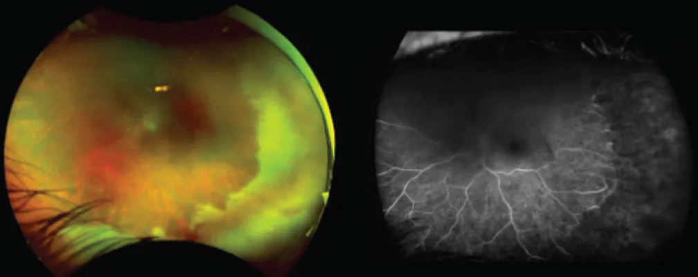
The patient underwent prompt treatment with intravitreal foscarnet and oral valacyclovir 1 g three times daily. Despite successful stabilization of the retinitis and serial anterior chamber taps, which remained negative for viral DNA, the patient developed a prolonged course of recurrent cystoid macular edema (CME) over the next 2 years.
CLINICAL COURSE
Following induction therapy, the patient’s retinitis regressed, and visual acuity initially stabilized. At month 3, the patient’s anterior chamber tap was negative for VZV, and systemic antiviral therapy was reduced to valacyclovir 1 g daily. However, over the ensuing months, she experienced recurrent and progressive CME with fluctuating visual acuity.
At month 14, there was concern for recurrent inflammation due to rare cell in the anterior chamber. In addition, we noted worsening CME while on topical prednisolone acetate and ketorolac, and her VA declined to 20/100. The anterior chamber tap was still negative for VZV. By month 20, we noted progressive CME and cataract formation. The patient remained on prophylactic valacyclovir 1 g daily during the entire clinical course after month 3; meanwhile, her VA declined again to 20/200, while the anterior chamber tap remained negative for VZV.
Here, we faced a therapeutic dilemma: Was the persistent CME inflammatory, infectious, ischemic, or multifactorial?
UNDERSTANDING CME IN ARN
CME after ARN is a complex and multifactorial process, with various pathogenic mechanisms. Caution is warranted when considering local steroids, as cases of viral reactivation have been reported with unopposed local steroids.1
The differential diagnosis of CME in ARN includes:
- Inflammatory CME: Persistent inflammation and blood-retinal barrier breakdown are common contributors to CME following ARN. Even after viral control is achieved, chronic immune activation can sustain vascular leakage and edema.2
- Infectious reactivation: Reactivation of VZV remains a serious concern for any patient with recurrent inflammation or worsening CME. Maintaining patients on a prophylactic regimen of systemic antiviral therapy is critical long-term, and treatment doses can be reintroduced when there is a concern for reactivation. Repeat aqueous sampling may help guide management decisions, particularly before escalating corticosteroid therapy.
- Ischemic and VEGF-driven CME: Peripheral retinal ischemia after necrotizing retinitis can induce VEGF upregulation and chronic vascular permeability. In these cases, VEGF-mediated edema may persist despite corticosteroid or antiviral therapy.3
Management options for CME following ARN include systemic, periocular, or intravitreal corticosteroids; continued antiviral suppression; intravitreal antiviral injection; intravitreal anti-VEGF injection; and other immunomodulatory agents (eg, tocilizumab or interferon alpha-2a).
In this patient, repeat negative aqueous taps and the absence of clinical signs of inflammation reduced our concern for active viral replication and shifted our attention toward inflammatory and ischemic mechanisms.
ANTI-VEGF THERAPY AS A TURNING POINT
At month 20, the patient received intravitreal aflibercept 8 mg (Eylea HD, Regeneron). Two weeks later, her VA improved from 20/200 to 20/60 with significant reduction in CME (Figure 2). Subsequently, the patient received intravitreal bevacizumab (Avastin, Genentech/Roche), sub-Tenon triamcinolone 40 mg, and cataract extraction with IOL placement. By month 27, her VA improved significantly to 20/25 with substantial restoration of macular architecture. Her vision remained stable without CME recurrence (Figure 3).
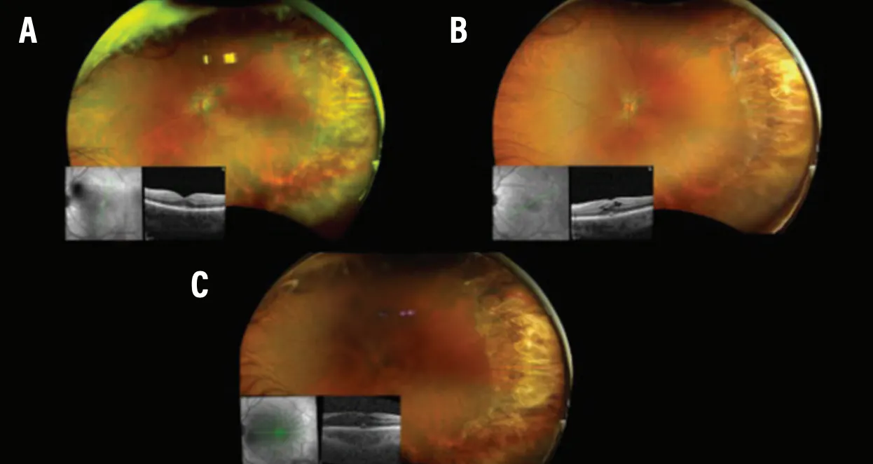
Figure 2. OCT at month 3 demonstrated chorioretinal scarring in the previously affected area without CME (A). At month 14, with no reactivation of ARN, OCT showed new CME (B). At month 20, OCT showed worsening posterior subcapsular cataract and CME despite topical ketorolac and prednisolone acetate (C).
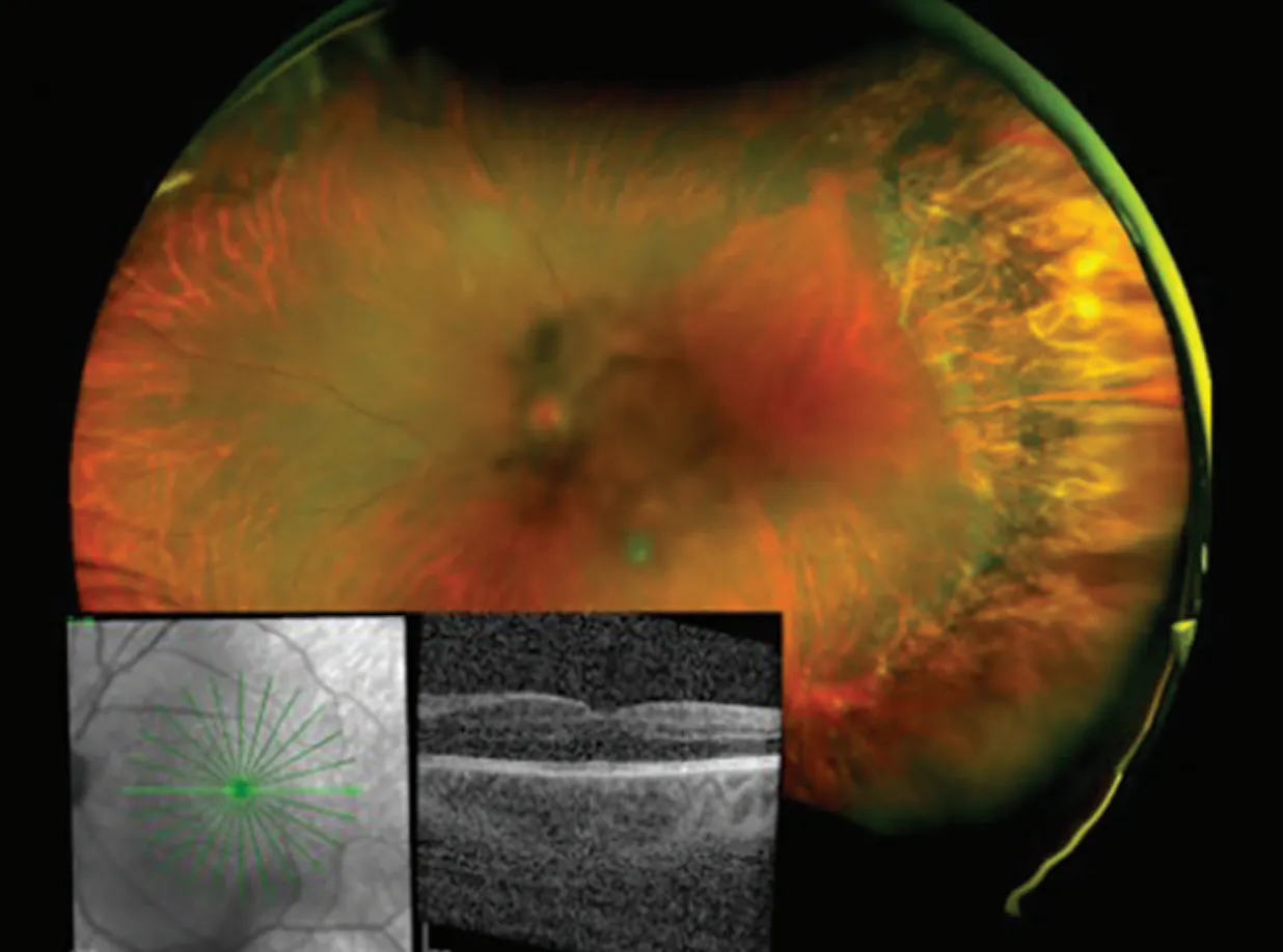
Figure 3. Two weeks after the initiation of intravitreal anti-VEGF therapy, OCT demonstrated resolution of CME.
This case highlights several important lessons in the management of chronic CME after ARN, including:
- CME in ARN is often multifactorial. Although inflammation is typically emphasized, VEGF-driven vascular permeability from peripheral ischemia may play a substantial role in persistent edema.
- Repeat viral testing can guide steroid escalation. In this patient, serial negative aqueous taps helped support escalation of antiinflammatory therapy, while reducing concern for active viral replication.
- Anti-VEGF therapy may be underused. The patient’s dramatic response to anti-VEGF therapy suggests VEGF-mediated vascular dysfunction may be an important therapeutic target in select patients with chronic CME after ARN.
- Long-term management requires flexibility. Patients with ARN frequently require individualized and evolving treatment over months to years. The balance between antiviral coverage, inflammation control, and management of ischemic complications can be delicate. While some patients may wean off antivirals, it is important to maintain prophylactic dosing in the setting of chronic inflammation, especially with local steroid therapy.
A MULTIFACTORIAL DISEAESE PROCESS
Chronic CME after ARN remains a challenge with limited evidence-based guidance. This case demonstrates how persistent edema may reflect overlapping inflammatory and ischemic mechanisms even after apparent viral quiescence.
Clinicians must maintain a broad differential diagnosis, using repeat viral testing when appropriate and considering anti-VEGF therapy in refractory cases. In select patients, targeting VEGF-mediated vascular leakage may lead to meaningful anatomic and visual recovery.
1. Chang S, Weissgold DJ, Singer JA, Sobrin L. Acute retinal necrosis after intraocular triamcinolone acetonide injection. Retin Cases Brief Rep. 2010;4(4):306-308. doi.org/10.1097/ICB.0b013e3181b5ee58
2. Wu P, Xing X, Huan Y, et al. Intravitreal dexamethasone implants for macular edema secondary to acute retinal necrosis. Ocul Immunol Inflamm. 2025;33(5):736-742. doi.org/10.1080/09273948.2024.2413692
3. Ortega-Evangelio L, Navarrete-Sanchis J, Williams BK, Tomás-Torrent JM. Aflibercept in the management of acute retinal necrosis syndrome-related macular edema. Eur J Ophthalmol. 2018;28(2):259-261. doi.org/10.5301/ejo.5001039


