




KEY TAKEAWAYS
- Central serous chorioretinopathy (CSCR) is a pachychoroid spectrum of disease characterized by localized serous detachments of the neurosensory retina due to increased choroidal vessel permeability.
- The authors report a case of a patient who developed sudden distortion of vision and a black spot in the central visual field of his right eye 6 days after his sinus surgery. He was ultimately diagnosed with CSCR.
- If CSCR does not spontaneously resolve after 4 to 6 months, treatment options include focal laser photocoagulation and photodynamic therapy.
Central serous chorioretinopathy (CSCR) is a pachychoroid spectrum of disease characterized by localized serous detachments of the neurosensory retina due to increased choroidal vessel permeability.1 The condition typically affects middle-aged men; the annual incidence rate of CSCR in men is one per 10,000. Although the pathophysiology of CSCR is unclear, it is associated with stress, elevated corticosteroid levels, and a hyperactive sympathetic nervous system.2 There is a hypothetical framework of venous overload choroidopathy, which explains a new concept in CSCR: Choroidal venous overload results in remodeling and intervortex venous anastomoses.3
CSCR IN A YOUNGER PATIENT
A 27-year-old man presented to our ophthalmology clinic with distorted vision in his right eye. He reported no history of diabetes, hypertension, or cardiac disease, nor any history of drug allergy, smoking, alcohol, antipsychotic medication, sleep disorders, chronic dyspepsia, stress, or steroid intake.
The patient informed us that he had undergone functional endoscopic sinus surgery (FESS) 1 week prior for nasal polyposis and chronic pansinusitis. His surgical records indicated the proper steps, including bilateral nasal polypectomy, maxillary antrostomy, uncinectomy, and anterior ethmoidectomy under general anesthesia.
Six days after his sinus surgery, he developed sudden distortion of vision and a black spot in the central visual field of his right eye. His VA was 20/30 OU. Slit-lamp examination revealed a normal anterior segment in each eye. The retinal examination showed a large serous detachment in his right macula without hemorrhage and extrafoveal retinal pigment epithelium (RPE) changes in his left eye (Figure 1). OCT of his right eye showed a subfoveal neurosensory retinal detachment; the left eye showed a small area of pigment epithelium detachment nasal to the fovea (Figure 2). Based on these findings, he was diagnosed with CSCR of his right eye.
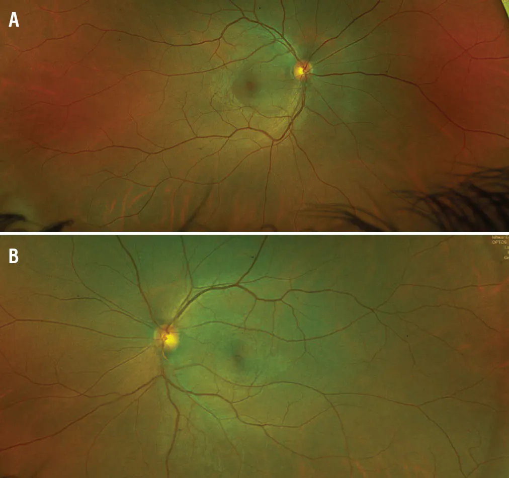
Figure 1. Fundus imaging of the right eye (A) showed serous macular detachment, while the left eye (B) showed a few RPE alterations.
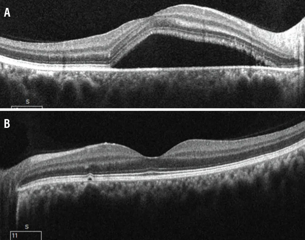
Figure 2. OCT of the right eye (A) showed serous subfoveal neurosensory detachment, while the left eye (B) showed a small pigment epithelium detachment nasal to the fovea.
His symptoms began to resolve gradually with observation. Full recovery of vision and resolution of symptoms were achieved 1 month after surgery.
PEARLS FOR POSTOPERATIVE CSCR
Theories explaining the pathophysiology of CSCR suggest that corticosteroids and epinephrine can potentiate choroidal vasoconstriction and relative ischemia, which can then lead to hyperpermeability in the choroidal vasculature as a result of changes in oncotic pressure and disruption of the RPE pump.4
To our knowledge, there have been no prior reports of this complication following FESS. In our case, there was no use of corticosteroids during the procedure or postoperatively. However, the surgeon used a 1:1000 epinephrine-soaked cotton swab intraoperatively during nasal packing.
CSCR may develop after FESS due to psychological stress associated with pain or postoperative difficulty; epinephrine used during nasal packing (ie, retrograde flow of epinephrine into the sinus, traveling backward through systemic valveless venous or arterial circulation, and entering the retinal artery); or sudden onset of orbital hematoma from anterior ethmoidectomy.
In our case, psychological stress or epinephrine use during the procedure could be possible causes for the development of CSCR. CSCR has also been reported to occur after rhinoplasty, orbital blowout fracture, or lacrimal surgery.
If the disease does not spontaneously resolve after 4 to 6 months, treatment options include focal laser photocoagulation and photodynamic therapy.5 Ear, nose, and throat surgeons should be aware that CSCR can occur as a result of FESS or epinephrine use during nasal packing. If CSCR is suspected in a patient after FESS, corticosteroids should be discontinued.
AWARNESS OF FESS
In this case, intraoperative use of topical epinephrine during nasal packing may have increased the risk of CSCR development. Topical spray containing another vasoconstrictor can be used before nasal packing. To prevent this potentially serious complication, efforts should also be made to alleviate any psychological stress.
1. Zhang X, Lim C, Chhablani J, Wong YM. Central serous chorioretinopathy: updates in the pathogenesis, diagnosis and therapeutic strategies. Eye Vis (Lond). 2023;10(1):33. doi.org/10.1186/s40662-023-00349-y
2. Mansour AM, Koaik M, Lima LH, et al. Physiologic and psychologic risk factors in central serous chorioretinopathy. Ophthalmol Retina. 2017;1(6):497-507. doi.org/10.1016/j.oret.2017.02.009
3. Zarnegar A, Ong J, Singh SR, Chhablani J. Venous overload choroidopathy. In: Retinal and Choroidal Vascular Diseases of the Eye. 2024;273-281.
4. Zarnegar A, Ong J, Matsyaraja T, Arora S, Chhablani J. Pathomechanisms in central serous chorioretinopathy: a recent update. Int J Retina Vitreous. 2023;9(1):3. doi.org/10.1186/s40942-023-00443-2
5. Shin YI, Kim KM, Lee MW, Kim JY, Jo YJ. Long-term results of focal laser photocoagulation and photodynamic therapy for the treatment of central serous chorioretinopathy. Jpn J Ophthalmol. 2019;64(1):28-36. doi.org/10.1007/s10384-019-00697-0


