




KEY TAKEAWAYS
- For surgeons managing proliferative vitreoretinopathy (PVR) following rhegmatogenous retinal detachment (RRD) repair, the challenge is not simply to reattach the retina, but to protect the macula at every step.
- Internal limiting membrane (ILM) peeling has been shown to significantly improve single-surgery anatomic success compared with no ILM peeling and reduce the need for persistent silicone oil as well as the rate of secondary epiretinal membrane surgery.
- Heavy silicone oil is a useful adjunct for inferior PVR-RRD, as it provides a superior tamponade to the inferior retina versus standard silicone oil and negates the need for strict postoperative posturing.
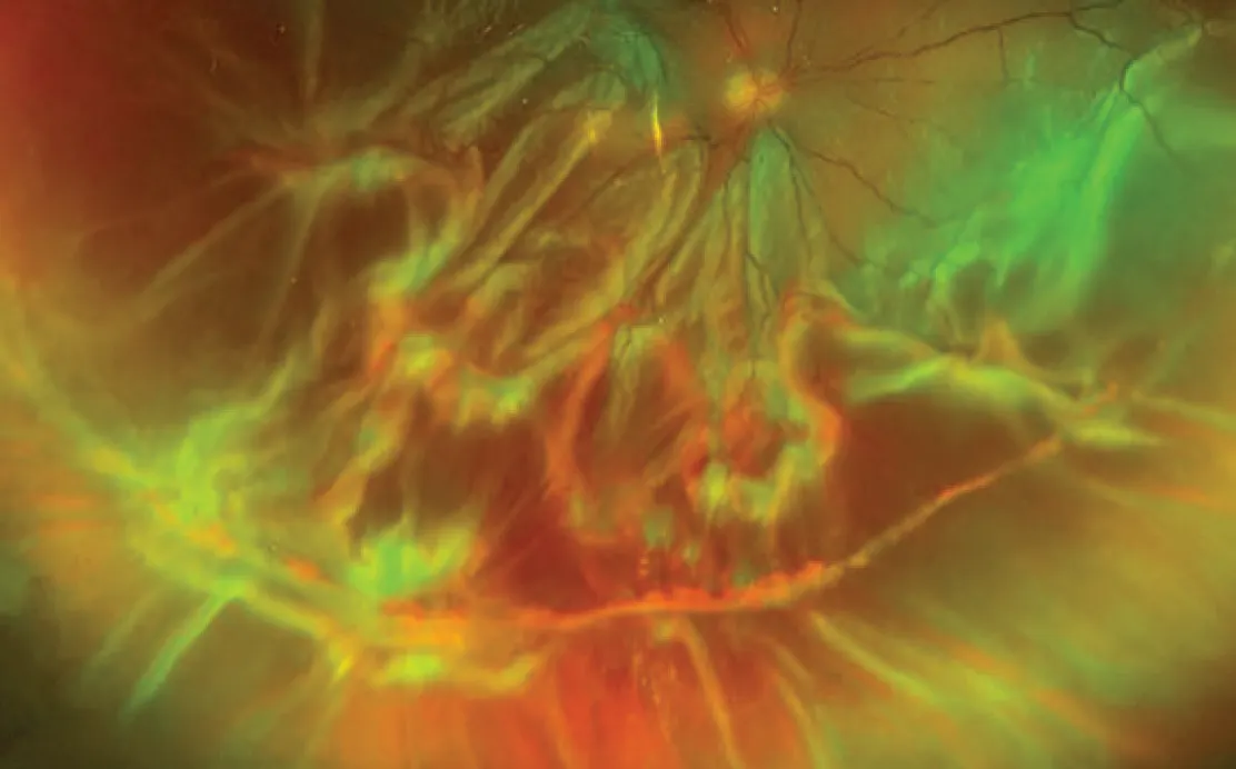
Proliferative vitreoretinopathy (PVR) remains a leading cause of surgical complications in rhegmatogenous retinal detachment (RRD) repair and an independent risk factor for poor visual prognosis (Figure 1).1,2 Even if anatomic reattachment is achieved, visual recovery often lags, limited by complications such as macular edema (ME), epiretinal membrane (ERM), macular folds, and photoreceptor loss from recurrent detachment.3,4 For vitreoretinal surgeons, the challenge is not simply to reattach the retina, but to protect the macula at every step, from preoperative imaging to tamponade selection and removal.
This article details practical, evidence-based strategies to optimize macular outcomes in PVR-RRD.
MACULAR BIOMARKERS OF VISUAL PROGNOSIS
Preoperative macular status should be documented with OCT and should be mandatory in the postoperative period. Subfoveal fluid, photoreceptor integrity and ERM or ME guide patient counselling and predict recovery.5,6
OCT biomarkers play an increasingly important role in predicting visual outcomes. In a post hoc longitudinal analysis of a randomized controlled trial of patients who underwent surgery for PVR-C RRD, early postoperative OCT findings, including retinal pigment epithelium and outer retinal layer thickness as well as qualitative markers (eg, gross retinal layer disruption, absence of foveal bulge) were significantly associated with poorer visual outcomes.7
Patient-reported outcome data add useful perspectives. Baseline and early postoperative BCVA are among the strongest predictors of 12-month visual function and vision-related quality of life.2,8 In PVR-RRD cohorts, final BCVA correlates most strongly with Visual Function Questionnaire-25 and Short Form Health Survey-36 scores, rather than the number of surgeries or anatomic reattachment rate.8
INTRAOPERATIVE MACULAR STRATEGY: TIPS AND TRICKS
Broad Internal Limiting Membrane (ILM) Peeling
In a large multicenter analysis (370 eyes with PVR-C RRD), ILM peeling1:
- significantly improved single-surgery anatomic success compared with no peeling at 3 months (86.6% vs 73.2%) and 6 months (75.2% vs 65.3%);
- raised the retina-attached-under-fluid rate at the final follow-up visit (84.7% vs 75.6%);
- reduced the need for persistent silicone oil; and
- halved the rate of secondary ERM surgery (8.9% vs 17.8%).1
In addition, extended ILM peeling beyond the vascular arcades was independently associated with better final visual acuity and with reattachment without oil tamponade.1 The rationale for these results is both mechanical and biological. The ILM is the scaffold on which proliferative membranes grow; removing it denies recurrent PVR its substrate.1,9 Peeling also confirms complete removal of overlying preretinal tissue and disconnects peripheral traction from the posterior pole.1,10
Practical Tips
- Stain with brilliant blue G or indocyanine green.
- Peeling under perfluorocarbon liquid (PFCL) is a good option when the retina is mobile; approximately 60% of ILM peels in the multicenter cohort used PFCL for counter-traction and improved visualization of the peeled edge.1
- Extend peeling as close as possible to any retinectomy margin (Figure 2), as that edge is the most common site of PVR proliferation and stretched breaks.1
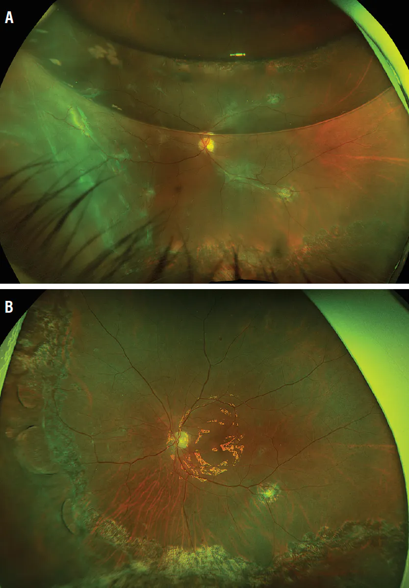
Figure 2. Preoperative fundus photography shows PVR redetachment under gas tamponade (A). Postoperatively, the retina is attached following ILM peeling, retinectomy, and silicone oil tamponade (B).
Redetachments in eyes without ILM peeling frequently involve posterior breaks, including macular holes from tangential contraction.1 Broad ILM peeling reduces this risk by eliminating the posterior scaffold. When a fold is identified intraoperatively, PFCL-assisted flattening, meticulous removal of the posterior hyaloid and epiretinal tissue, and ILM peeling across the fold minimize persistent distortion.
Subretinal PVR
Subretinal bands are part of the PVR-C spectrum and contribute to posterior tractional tethering.11 They should be removed only when they are clearly preventing reattachment, while small, non-contracting strands can be left alone if the retina flattens. When extensive, a nonmacular retinotomy is preferable to risking direct foveal manipulation.
CHOOSING THE RIGHT TAMPONADE
Tamponade with silicone oil or a long-acting gas (eg, C3F8) remains the standard approach for managing PVR. The Silicone Study demonstrated broadly comparable anatomic outcomes between the two types of tamponades for PVR-C RRD cases, while shorter-acting SF6 has consistently been associated with higher redetachment risk.12,13
Silicone oil remains the most common tamponade for PVR-C but is not benign to the macula. It can induce ME through both macrophage-driven inflammation and mechanical traction at the oil-retina interface.14 Moreover, longer oil duration correlates with worse visual outcomes.2,15 To facilitate earlier silicone oil removal, peel the ILM up to the retinectomy margin to reduce PVR proliferation and stretched breaks, thus protecting the macula.1
Heavy silicone oil is also a useful adjunct for inferior PVR-RRD, as it provides a superior tamponade to the inferior retina versus standard silicone oil and negates the need for strict posture.16 It should also be removed early (approximately 8 weeks postoperatively) and requires close monitoring for IOP and lens-related complications in the postoperative period.17
Gas tamponade should be considered in cases with a low risk of reproliferation, no active inflammation, and in which membranes have been fully removed.
MANAGEMENT OF MACULAR CONSEQUENCES AND EDEMA
Postoperative increase in ERM and cystoid ME (CME), along with early retinal layer disruptions, are important predictors of long-term visual outcomes.
Disrupted ellipsoid zone and external limiting membrane can be associated with poorer visual outcomes, supporting a hypothesis that external limiting membrane disruption may represent a shift from a protective to a detrimental glial response.15,18,19
Postoperative CME should be approached with a stepwise, inflammation-focused strategy. Topical nonsteroidal antiinflammatory drugs or steroids alone or in combination remain first-line therapies.4
For recalcitrant or chronic ME, an intravitreal dexamethasone implant (Ozurdex, Abbvie) is an option; in a randomized trial of PVR-C, a dexamethasone implant reduced 6-month CME incidence versus placebo (42.7% vs. 67.2%).19
However, ME recurrence is common, and most eyes require repeat implantation. Early intervention (within 4 months of ME diagnosis) has been found to reduce the total number of injections needed.4,20 IOP elevation and cataract progression are the main risks and must be monitored.4 ERM formation is strongly linked to persistent ME, and removal is likely to improve visual outcomes.
IMPROVE OUTCOMES WITH PVR
The trends in PVR surgery are clear: more complete mechanical removal of inflammatory scaffolds, earlier antiinflammatory pharmacotherapy, and shorter silicone oil duration. As we move forward, revision of the current classification of PVR is necessary to standardize care, provide a decision-making guide, identify high-risk cases, and improve outcomes. Large, randomized clinical trials are required to assess the effectiveness of adjunctive treatment options in combination with surgery for the management of PVR.
1. Wakabayashi T, Samuelson AG, Oshima Y, et al. Internal limiting membrane peeling for grade C proliferative vitreoretinopathy: an international multicenter study. Ophthalmol Retina. 2026;10(1):5-16. doi.org/10.1016/j.oret.2025.06.006
2. Wickham L, Ho-Yen GO, Bunce C, et al. Surgical failure following primary retinal detachment surgery by vitrectomy: risk factors and functional outcomes. Br J Ophthalmol. 2011;95(9):1234-1238. doi.org/10.1136/bjo.2010.190306
3. Pastor JC, Rojas J, Pastor-Idoate S, et al. Proliferative vitreoretinopathy: A new concept of disease pathogenesis and practical consequences. Prog Retin Eye Res. 2016;51:125-155. doi.org/10.1016/j.preteyeres.2015.07.005
4. Ferro Desideri L, Arun K, Melo GB, et al. Macular edema after rhegmatogenous retinal detachment: can we treat it effectively? Exp Rev Ophthalmol. 2024;19(6):401-404. doi.org/10.1080/17469899.2024.2390435
5. Pole C, Chehaibou I, Govetto A, et al. Macular edema after rhegmatogenous retinal detachment repair: risk factors, OCT analysis, and treatment responses. Int J Retina Vitreous. 2021;7(1):9. doi.org/10.1186/s40942-020-00254-9
6. Ferro Desideri L, Danilovska T, Bernardi E, et al. Artificial intelligence-enhanced OCT biomarkers analysis in macula-off rhegmatogenous retinal detachment patients. Transl Vis Sci Technol. 2024;13(10):21. doi.org/10.1167/tvst.13.10.21
7. Anguita Rodrigo SPY, Lorenzo FD, Moritz S, Philip B, Martin Z, David GC. Macular structure and function in retinal detachment complicated by proliferative vitreoretinopathy. Manuscript under review.
8. Anguita R, Ferro Desideri L, Roth J, et al. Patient-reported outcomes after surgery for proliferative vitreoretinopathy: post hoc analysis of a randomized controlled trial. Ophthalmologica. 2026;249(2):175-181. doi.org/10.1159/000550679
9. Forlini M, Date P, Ferrari LM, et al. Comparative analysis of retinal reattachment surgery with or without internal limiting membrane peeling to prevent postoperative macular pucker. Retina. 2018;38(9):1770-1776. doi.org/10.1097/IAE.0000000000001775
10. Francone A, Charles M. Extensive internal limiting membrane peeling for proliferative vitreoretinopathy. Int Ophthalmol. 2023;43(1):147-153. doi.org/10.1007/s10792-022-02397-0
11. Machemer R, Aaberg TM, Freeman HM, et al. An updated classification of retinal detachment with proliferative vitreoretinopathy. Am J Ophthalmol. 1991;112(2):159-165. doi.org/10.1016/s0002-9394(14)76695-4
12. Martha Faraby VA, Elvioza, Djatikusumo Ari, Andayani Gitalisa, Yudantha Anggun. Various tamponade in surgery for retinal detachment associated with proliferative vitreoretinopathy. Journal of the Indonesian Opthalmologist Association. 2015;41(2):131-145. doi.org/10.35749/journal.v41i2.24
13. Abrams GW, Azen SP, McCuen BW 2nd, et al. Vitrectomy with silicone oil or long-acting gas in eyes with severe proliferative vitreoretinopathy: results of additional and long-term follow-up: Silicone Study report 11. Arch Ophthalmol. 1997;115(3):335-344. doi.org/10.1001/archopht.1997.01100150337005
14. Kontou EP, Karakosta C, Kounas K, et al. Macular edema following silicone oil tamponade for retinal detachment: a literature review. Cureus. 2023;15(12):e51233. doi.org/10.7759/cureus.51233
15. Quiram PA, Gonzales CR, Hu W, et al. Outcomes of vitrectomy with inferior retinectomy in patients with recurrent rhegmatogenous retinal detachments and proliferative vitreoretinopathy. Ophthalmology. 2006;113(11):2041-2047. doi.org/10.1016/j.ophtha.2006.05.039
16. Liu F, Li H, Feng L, et al. Anatomical and functional outcomes after Densiron 68 heavy silicone oil tamponade for complicated retinal detachment in Chinese eyes. Int J Ophthalmol. 2014;7(3):469-473. doi.org/10.3980/j.issn.2222-3959.2014.03.15
17. Hammer M, Ie A, Eibenberger K, et al. An analysis of heavy silicone oil treatment for inferior proliferative vitreoretinopathy. BMC Ophthalmol. 2025;25(1):38. doi.org/10.1186/s12886-024-03834-7
18. Kon CH, Occleston NL, Aylward GW, et al. Expression of vitreous cytokines in proliferative vitreoretinopathy: a prospective study. Invest Ophthalmol Vis Sci. 1999;40(3):705-712.
19. Banerjee PJ, Quartilho A, Bunce C, et al. Slow-release dexamethasone in proliferative vitreoretinopathy: a prospective, randomized controlled clinical trial. Ophthalmology. 2017;124(6):757-767. doi.org/10.1016/j.ophtha.2017.01.021
20. Thanos A, Todorich B, Yonekawa Y, et al. Dexamethasone intravitreal implant for the treatment of recalcitrant macular edema after rhegmatogenous retinal detachment repair. Retina. 2018;38(6):1084-1090. doi.org/10.1097/IAE.0000000000001720


