





KEY TAKEAWAYS
- Marfan syndrome (MFS) is a genetic disease affecting connective tissue with autosomal dominant transmission; broad involvement occurs, including cardiovascular, musculoskeletal, dermatological, and ocular complications.
- The authors report a case of a 34-year-old woman with MFS who developed elevated, uncontrolled IOP following 23-gauge PPV for the repair of a retinal detachment in her left eye and a 360° prophylactic laser retinopexy in her right eye.
- The authors propose that a scleral patch graft may have been more appropriate for closing the compromised thin sclera in this patient with MFS.
Marfan syndrome (MFS) is an autosomal dominant genetic disease affecting connective tissue. The defect lies in the FBN1 gene on chromosome 15, encoding fibrillin, an extracellular matrix glycoprotein.1,2 Broad involvement occurs in MFS, notably with cardiovascular, musculoskeletal, dermatological, and ocular effects.3,4 Ocular manifestations include ectopia lentis, microspherophakia, myopia, glaucoma, retinal tears, and retinal detachment (RD).5
In this article, we describe a rare case of scleral dehiscence following 23-gauge pars plana vitrectomy (PPV) for RD repair in a patient with MFS. This case highlights the potential effects of MFS on the scleral tissue following PPV, identifies contributing factors, and offers ways to anticipate them for future surgical interventions.
CASE REPORT
A 34-year-old woman with MFS presented with a sudden decrease in vision in her left eye. She had undergone intracapsular lens extraction and iris clip IOL implantation for bilateral crystalline lens subluxation 2 years earlier. On examination, her BCVA was 20/20 OD and hand motion OS. She was highly myopic with an axial length of 28.72 mm OD and 27.59 mm OS. Her IOP was 14 mm Hg OD and 11 mm Hg OS. She had corneal scarring and localized iris atrophy from her previous anterior segment surgeries. The iris clip IOL was slightly off-center inferiorly in each eye (Figure 1).
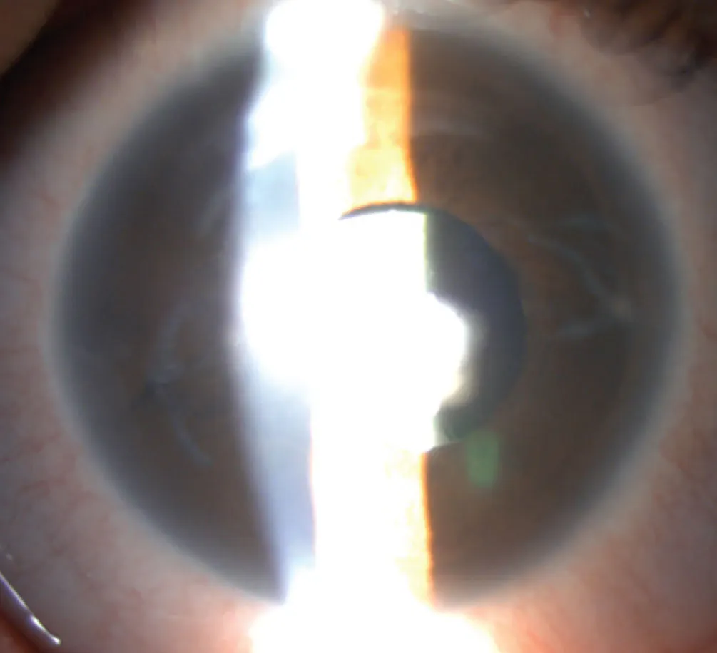
Figure 1. This photograph shows corneal scarring, localized iris atrophy, and slight inferior decentration of the IOL in the left eye.
On fundoscopic examination, we found a total bullous RD in the left eye and a horseshoe tear at the 11:00 clock hour. The retina of the right eye showed signs of multiples peripheral degenerations in the form of palisades.
We opted for a 23-gauge PPV for the repair of the RD in her left eye and a 360° prophylactic laser retinopexy for the right eye. A three-port vitrectomy was performed without incident, and endolaser retinopexy was applied to barricade the tear and for 360° cerclage. C2F6 gas was used as an endotamponade with a bubble slipping into the anterior chamber at the moment of exchange. Finally, the sclerotomies were sutured with an 8-0 vicryl, as they were persistently leaking at the end of the surgery.
POSTOPERATIVE COMPLICATIONS
On postoperative day 1, the IOP increased to 45 mm Hg OS. The gas bubble that had slipped into the anterior chamber was pressing on the IOL, causing a narrowing of the inferior angle. The patient was prescribed IOP-lowering medication and instructed to remain in a face-down position to allow the gas bubble to regain the posterior segment. The IOP successfully returned to baseline. The patient was discharged 5 days after surgery, with the maintenance of IOP-lowering medications.
One week postoperatively, we unsuccessfully tried to taper the IOP-lowering medications. The gas bubble in the anterior chamber regressed, but the inferior angle remained narrow. At the 2-month follow-up, her VA was 20/200 OS. We noticed pigmented masses protruding through the sclera at the trocar insertion points (Figure 2). IOP was 42 mm Hg OS despite a combination of IOP-lowering drops.
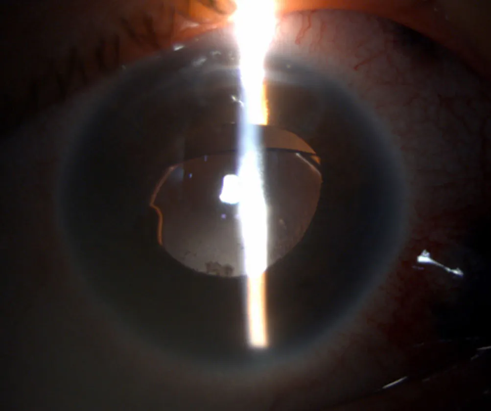
The anterior chamber was irregular with an inferiorly displaced IOL (Figure 3). On gonioscopy, the inferior angle was fully closed for more than 180° with some localized peripheral anterior synechiae. The retina remained flat.
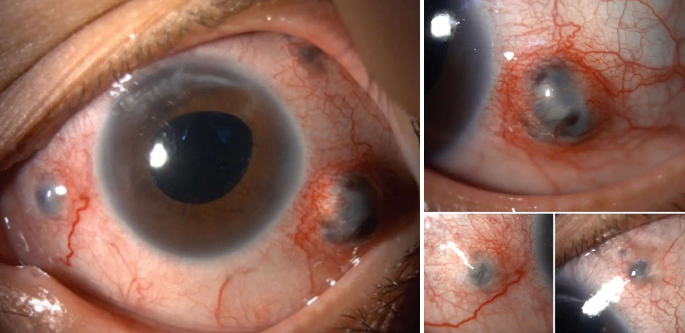
Figure 2. These photographs show the bulging of the choroid through the areas of scleral thinning at the trocar insertion points.
OCULAR COMPLICATIONS OF MFS
In the setting of ubiquitous pathological connective tissue in MFS, the sclera is also affected, where it is unusually thin and exhibits reduced resistance to mechanical stress. Scleral thinning and perforation can be precipitated by trauma, surgery, or even by centrifugal force emanating from within the eye, such as with elevated IOP.6,7 Extreme cases of spontaneous scleral rupture have been reported.8,9 Our patient’s case appears to combine multiple risk factors: The surgical insult complicated by increased IOP, which could explain the rapid progression of the choroidal bulge through the thinned sclera, instead of the late appearance described in the literature.10-12
Given the high risk of RD in MFS, surgeons must contend with pathological tissue that can compromise surgical outcomes and visual prognosis. Deramo et al reported a case of full-thickness scleral erosion in a 33-year-old man with MFS who underwent RD repair with a silicone sponge and silicone encircling band.7 PPV does not seem safer, as Sridhar et al reported a case of sclerotomy wound dehiscence after 20-gauge PPV and lensectomy for an ectopic lens in a 19-year-old woman.10 Similarly, Mancino et al reported a case of scleral wound dehiscence in a 34-year-old woman with MFS who underwent lensectomy 20 years prior.11 However, those cases relied on 20-gauge ports. With the advent of 23-, 25-, and 27-gauge ports, this complication is likely to be significantly reduced; at the time of our research, we found no cases in which scleral dehiscence occurred after a 23-gauge PPV for RD repair.
For repair options of scleral dehiscence, it can be closed with nylon sutures.7,10 Alternatively, it may require more advanced techniques with an autologous scleral graft or preserved scleral donor patch graft.8 Rodríguez-Ares et al described another technique that consists of combining a scleral homograft and amniotic membrane transplant as an alternative to autologous scleral and conjunctival grafts.6 Furthermore, Stanciu et al described a multilayer technique that involves first a direct suture with 10-0 vicryl, the placement of a scleral graft sutured with 8-0 vicryl, and a dried amniotic membrane glued on top.12
In our case, we recognize that a scleral patch graft might have been more appropriate for closing the compromised thin sclera in MFS. Furthermore, it was crucial to strictly control IOP to avoid exacerbating this complication in the postoperative period.
ALWAYS PLAN AHEAD
Vitreoretinal specialists must be aware of the risk of scleral dehiscence when treating RD in patients with MFS, even with the advent of new microincisional vitreoretinal surgery techniques. Furthermore, elevated and uncontrolled IOP can exacerbate surgical trauma in these cases, requiring meticulous postoperative management.
Acknowledgement: Informed consent was obtained from the patient to use the clinical data in this case report. The principles of the Declaration of Helsinki have been respected.
1. Ammash NM, Sundt TM, Connolly HM. Marfan syndrome-diagnosis and management. Curr Probl Cardiol. 2008;33(1):37-39. doi.org/10.1016/j.cpcardiol.2007.10.001
2. Gould RA, Sinha R, Aziz H, et al. Multi-scale biomechanical remodeling in aging and genetic mutant murine mitral valve leaflets: insights into Marfan syndrome. PLoS One. 2012;7(9):e44639. doi.org/10.1371/journal.pone.0044639
3. Demetracopoulos CA, Sponseller PD. Spinal deformities in Marfan syndrome. Orthop Clin North Am. 2007;38(4):563-572. doi.org/10.1016/j.ocl.2007.04.003
4. Dietz HC, Cutting GR, Pyeritz RE, et al. Marfan syndrome caused by a recurrent de novo missense mutation in the fibrillin gene. Nature. 1991;352(6333):337-339. doi.org/10.1038/352337a0
5. Nemet AY, Assia EI, Apple DJ, Barequet IS. Current concepts of ocular manifestations in Marfan syndrome. Surv Ophthalmol. 2006;51(6):561-575. doi.org/10.1016/j.survophthal.2006.08.008
6. Rodríguez-Ares MT, Touriño R, Capeans C, Sánchez-Salorio M. Repair of scleral perforation with preserved scleral and amniotic membrane in Marfan’s syndrome. Ophthalmic Surg Lasers. 1999;30(6):485-487.
7. Deramo VA, Haupert CL, Fekrat S, Postel EA. Hypotony caused by scleral buckle erosion in Marfan syndrome. Am J Ophthalmol. 2001;132(3):429-431. doi.org/10.1016/s0002-9394(01)00993-x
8. Turaga K, Senthil S, Jalali S. Recurrent spontaneous scleral rupture in Marfan’s syndrome. BMJ Case Rep. 2016;2016:bcr2016214764. doi.org/10.1136/bcr-2016-214764
9. Voulgari N, Giacuzzo C, Schalenbourg A, Kymionis GD. Occult spontaneous ocular perforation presenting as conjunctival chemosis in a patient with Marfan’s syndrome. Case Rep Ophthalmol. 2019;10(3):344-348. doi.org/10.1159/000503440
10. Sridhar J, Chang JS, Aziz HA, Erickson BP. Delayed sclerotomy wound dehiscence after lensectomy and vitrectomy in Marfan syndrome. Oman J Ophthalmol. 2015;8(3):198-199. doi.org/10.4103/0974-620X.169893
11. Mancino R, Aiello F, Ceccarelli S, et al. Autologous conjunctival epithelium transplantation and scleral patch graft for postlensectomy wound leakage in Marfan syndrome. Eur J Ophthalmol. 2012;22(5):830-833. doi.org/10.5301/ejo.5000124
12. Stanciu PE, O’Regan A, Cosgrave E. Late onset sclerotomy dehiscence in a patient with Marfan syndrome presenting as recurrent episodes of raised intraocular pressure. BMJ Case Rep. 2022;15(8):e249990. doi.org/10.1136/bcr-2022-249990


