


KEY TAKEAWAYS
- Despite their overall excellent safety profile, anti-VEGF injections can, rarely, lead to serious adverse effects.
- The authors present a case of a patient who developed a full-thickness macular hole (FTMH) following a course of anti-VEGF intravitreal injections for macular edema secondary to combined central retinal artery and vein occlusion.
- It is important to acknowledge that development of a FTMH is a known risk in patients with chronic macular edema, such as the one in this case.
Intravitreal anti-VEGF injections are a mainstay of treatment for many retinal conditions, including wet AMD, macular edema following retinal vein occlusion (RVO), diabetic macular edema, diabetic retinopathy, and myopic choroidal neovascularization.1,2 Despite their overall excellent safety profile, anti-VEGF injections can, rarely, lead to serious adverse effects, including endophthalmitis, rhegmatogenous retinal detachment, RVO, and others.2 Development of a full-thickness macular hole (FTMH) has also been described, which is likely secondary to the severe contraction of the neovascular membrane and focal vitreoretinal traction forces induced by the anti-VEGF agent.3-9
Here, we present our case of a patient who developed a FTMH following a course of anti-VEGF intravitreal injections for macular edema secondary to combined central retinal artery and vein occlusion (CCRAVO).
CASE EXAMPLE
A 54-year-old man presented with a 1-month history of vision loss in his right eye. His corrected distance VA (CDVA) was counting fingers at 2 m OD and 20/25 OS, and his IOP was 14 mm Hg OU. No abnormalities were observed in the anterior segment of either eye. Fundus examination of his left eye was normal, while his right eye revealed intraretinal hemorrhages and venous engorgement in all four quadrants demonstrating macular whitening (ischemic retinal edema) consistent with central retinal artery occlusion, as well as a “cherry-red spot” or foveal-sparing and peripheral intraretinal hemorrhages typical of central RVO (Figure 1). These findings were consistent with a diagnosis of CCRAVO.10
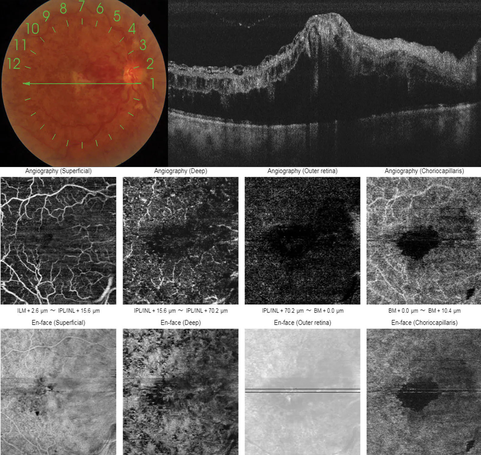
Figure 1. OCT and OCTA at baseline showing hyperreflective inner layers, subretinal fluid, and macular edema, with reduced vascular density in both the superficial and deep plexuses.
OCT angiography (OCTA) of the patient’s right eye showed macular edema, hyperreflective inner layers, and subretinal fluid, likely secondary to CCRAVO. He was treated with three monthly intravitreal injections of ranibizumab (Lucentis, Genentech/Roche).
One month after the third injection, the patient’s CDVA was counting fingers at 50 cm in his right eye. OCTA showed a FTMH, macular edema at its border, atrophy of the inner retinal layers, and signs of retinal ischemia (Figure 2).
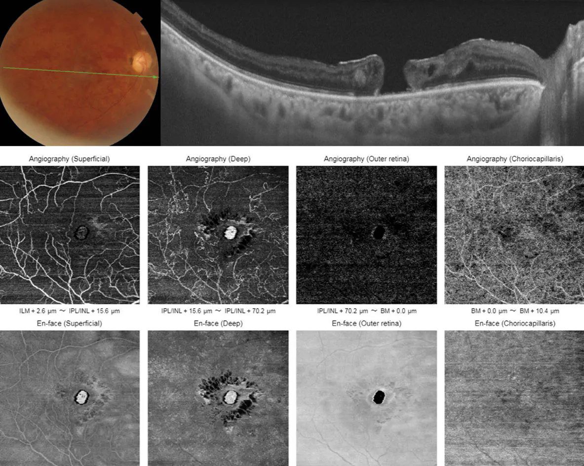
Figure 2. One month after the third intravitreal ranibizumab injection, OCT demonstrated an FTMH with persistent edema and inner retinal atrophy, while OCTA revealed vascular rarefaction and ischemic changes in the superficial and deep plexuses.
MANAGEMENT
Surgical repair via pars plana vitrectomy and internal limiting membrane peeling was deferred due to poor visual potential, extensive inner retinal atrophy, and profound macular ischemia as evidenced by OCTA. The presence of significant inner retinal atrophy, persistent macular edema, and signs of extensive ischemia further reduced the likelihood of functional visual recovery following vitrectomy.
Considering the widespread retinal ischemia observed on OCTA, the patient underwent panretinal laser photocoagulation in his right eye as a prophylactic measure to reduce the risk of complications, particularly neovascular glaucoma. He continues to be monitored regularly with multimodal imaging.
ABOUT CCRAVO
CCRAVO is a rare entity associated with multiple systemic diseases such as diabetes, lupus erythematosus, homocysteinemia, and dyslipidemia; therefore, management should involve a multidisciplinary approach.10 Despite treatment, visual prognosis remains poor.
VEGF has been found to be elevated in RVO, leading to persistent vascular leakage and macular edema. Encouraging results have been published regarding the efficacy of intravitreal anti-VEGF therapy on diminishing macular edema, halting neovascularization, and improving final visual acuity; complications, albeit rare, can occur, including infectious endophthalmitis, uveitis, rhegmatogenous retinal detachment, temporal IOP elevation, ocular hemorrhage, and very uncommonly, FTMH.2-4
Several mechanisms have been proposed to explain the presentation of FTMH after intravitreal anti-VEGF injection. Some authors have suggested the globe deformation and potential induction of vitreous incarceration in the injection site following the procedure could enhance vitreomacular traction (VMT), especially with an overlying pigment epithelial detachment (PED), subsequently leading to FTMH development.3,4 In addition, the chemical compounds introduced with the anti-VEGF therapy and the structural changes it induces could lead to VMT.5
In the available published cases of FTMH after intravitreal anti-VEGF injection,3-9 most FTMHs developed in patients whose indication for the procedure was wet AMD, with or without VMT or PED. In almost all cases, the FTMH diagnosis was confirmed by OCT and fluorescein angiography, apart from one study, in which only OCT was used.6 Surgical intervention led to improved visual outcomes but, unlike the case presented here, none of these reported cases had subjacent arterial occlusive compromise.
UNDERSTAND EVEN THE RAREST OF ADVERSE EVENTS
Intravitreal anti-VEGF injection plays an important role in the treatment of multiple retinal conditions. Although their most common adverse events are well known, FTMH has been reported in rare cases. It is important to acknowledge that development of a FTMH is a known risk in patients with chronic macular edema, such as the one in this case; thus, it is prudent to maintain a high degree of suspicion of this complication, particularly in eyes with severe ischemic compromise. Adequate follow-up with OCT can facilitate a prompt diagnosis and treatment, if indicated, to improve final visual acuity.
1. Sanchez JG, Ocampo HH, Rodriguez FJ, Grisales MA, Abdala C, Ruiz AJ. Anti-VEGF therapy for retinal diseases treatment: recommendations for its practice in Colombia. Rev Soc Colomb Oftalmol. 2018;51:46-62.
2. Ghasemi Falavarjani K, Nguyen Q. Adverse events and complications associated with intravitreal injection of anti-VEGF agents: a review of literature. Eye (Lond). 2013;27(7):787-794.
3. Kabanarou S, Xirou T, Mangouritsas G, et al. Full-thickness macular hole formation following anti-VEGF injections for neovascular age-related macular degeneration. Clin Interv Aging. 2017;12:911-915.
4. Querques G, Souied E, Soubrane G. Macular hole following intravitreal ranibizumab injection for choroidal neovascular membrane caused by age-related macular degeneration. Acta Ophthalmol. 2009;87(2):235-237.
5. Raiji V, Eliott D, Sadda S. Macular hole overlying pigment epithelial detachment after intravitreal injection with ranibizumab. Retin Cases Brief Rep. 2013;7(1):91-94.
6. Mukherjee C, Mitra A, Kumar N, Elsherbiny S, Lip P. Macular hole formation after intravitreal ranibizumab injection in wet age-related macular degeneration. Open Ophthalmol J. 2015;9:177-180.
7. Muramatsu D, Mitsuhashi R, Iwasaki T, Goto H, Miura M. Macular hole formation following intravitreal injection of ranibizumab for branch retinal vein occlusion: a case report. BMC Res Notes. 2015;8:358.
8. Goel N. Full-thickness macular hole formation and spontaneous closure during intravitreal ranibizumab therapy for central retinal vein occlusion. Retin Cases Brief Rep. 2022;16(6):678-680.
9. Moll-Udina A, Alforja-Castiella S, Figueroa-Vercellino JP, Alé-Chilet A, Casaroli-Marano RP. Simultaneous retinal pigment epithelium tear and lamellar macular hole evolving to a full-thickness macular hole after intravitreal injection. J Fr Ophtalmol. 2020;43(7):e237-e239.
10. Sengupta S, Pan U. Combined branch retinal vein and branch retinal artery occlusion - clinical features, systemic associations, and outcomes. Indian J Ophthalmol. 2017;65(3):238-241.

_1784132761.jpg?auto=compress,format&w=70)


