


Corneal size has a significant influence on planning in refractive surgery, particularly in cases of microcornea. Microcornea is associated with congenital cataracts, predisposing patients to a greater risk of cataractous change in early adulthood.1 This condition can appear alone or with microphthalmos. Microphthalmos can be associated with systemic abnormalities and is frequently accompanied by congenital cataract and nystagmus.2
Such anatomic abnormalities present unique surgical challenges that affect incision size, technique for cataract extraction, IOL selection, power calculation, and implantation1,3; consequently, a specialized technique is often required. Furthermore, aphakic spectacles can induce significant optical aberration, which is often more pronounced with nystagmus.
Here, we report a case of a patient with bilateral aphakia, microcornea, nystagmus, and vitreous opacity who underwent pars plana vitrectomy (PPV) and secondary IOL implantation in each eye using a modified Yamane technique featuring custom haptic trimming without glued scleral fixation, resulting in improved visual outcomes.
CASE REPORT
A 28-year-old woman with bilateral aphakia, microcornea, nystagmus, and vitreous opacity presented to our hospital seeking refractive correction. She had a history of bilateral cataract surgery performed in childhood, which resulted in aphakia in each eye. A comprehensive preoperative evaluation was performed, including visual acuity assessment, refraction, tonometry, slit-lamp biomicroscopy, and OCT of the anterior segment and macula.
Her preoperative uncorrected VA (UCVA) was 20/600 OU, improving to 20/200 OU with pinhole. Axial length measurements were 21.86 mm OD and 20.66 mm OS. This revealed a significant interocular asymmetry of 1.2 mm, far exceeding the normal physiological variation (< 0.3 mm) and consistent with a diagnosis of asymmetric bilateral microphthalmos. Her horizontal corneal diameters were 10 mm OD and 9.9 mm OS, confirming the diagnosis of bilateral microcornea.
Keratometry readings were obtained as part of the ocular biometry. The values were K1 41.88 D and K2 44.20 D in her right eye and K1 41.61 D and K2 43.83 D in her left eye, which fell within the normal physiological range (approximately 42-45 D). The resulting keratometric astigmatism was -2.32 D at 2° in her right eye and -2.22 D at 30° in her left eye, indicating bilateral, moderate, with-the-rule astigmatism. The presence of a regular astigmatic pattern was a favorable finding, suggesting a relatively well-formed corneal surface despite the reduced diameter and allowing for a predictable optical correction.
SURGICAL APPROACH
Considering the patient’s bilateral aphakia, significant vitreous opacity, and nystagmus, the decision was made to proceed with PPV and secondary IOL implantation under general anesthesia. The first procedure was performed on her right eye, and after postoperative follow-up showed no complications, we operated on her left eye 1 month later.
A monofocal, acrylic, three-piece IOL (optic diameter: 6.00 mm; haptic-to-haptic distance: 13 mm; power: +27.0 D OD and +26.0 D OS) was selected. A modified Yamane technique, which featured custom haptic trimming and omitted glued scleral fixation, was performed (Figure 1, Video). The steps of this procedure were as follows:
- Insert three 25-gauge vitrectomy trocars and perform a core vitrectomy to clear the vitreous opacities.
- Create a superior corneal incision using a keratome at the 12:00 clock position to allow injection of a viscoelastic substance and subsequent insertion of the IOL into the anterior chamber.
- Perform an anterior vitrectomy.
- Use scleral marks to indicate the intended fixation points for the haptics (right and left).
- Insert a 26-gauge needle obliquely at a 30° angle through the sclera approximately 2 mm from the limbus to externalize the leading IOL haptic.
- Trim the IOL haptic, withdrawn by approximately 1 mm, after which its tip is flanged to a diameter of 0.3 mm to 0.5 mm using low-temperature cautery to secure its fixation.
- Externalize the second haptic using another 26-gauge needle placed 180° from the first. It is then trimmed by 1 mm and flanged without additional glue or sutures required for IOL fixation.
- Perform irrigation and aspiration to remove the viscoelastic substance from the anterior chamber.
- Remove the trocar cannulas.
- Administer a subconjunctival injection of dexamethasone.
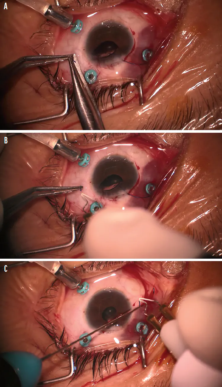
Figure 1. The first trimmed haptic (A) and low-temperature cautery (B). The second trimmed haptic with low-temperature cautery for fixation (C). Note the symmetrical shortening of both haptics and the rounded, flanged tip (0.3-0.5 mm) created by low-temperature cautery, which secures intrascleral fixation without glue or sutures.
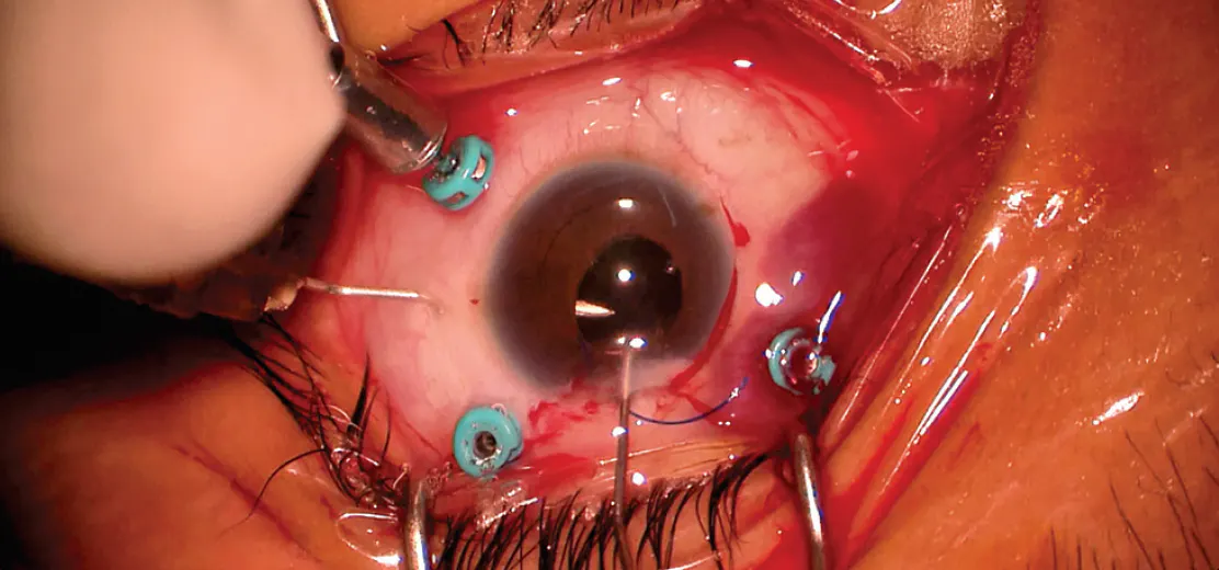
Video. A Modified Yamane Technique Featuring Custom Haptic Trimming Without Glued Scleral Fixation.
POSTOPERATIVE OUTCOMES
At the 1-month postoperative visit, the patient’s UCVA improved to 20/200 OU with BCVA of 20/100 OU. She also reported subjective improvement in vision and quality of life. At the 14-month postoperative evaluation, improvements in BCVA were maintained. We also performed detailed anterior segment analysis of each eye using anterior segment OCT and horizontal B-scan (Figures 2 and 3). The patient’s nystagmus made it difficult to obtain optimal OCT scan quality, which might have affected the precision of the quantitative measurements; however, we observed no obvious signs of macular edema, atrophy, or other structural abnormalities on qualitative assessment of the B-scans (Figure 3C and D). An in-depth postoperative OCT analysis of the optic nerve head and retinal nerve fiber layer revealed no abnormalities in either eye.
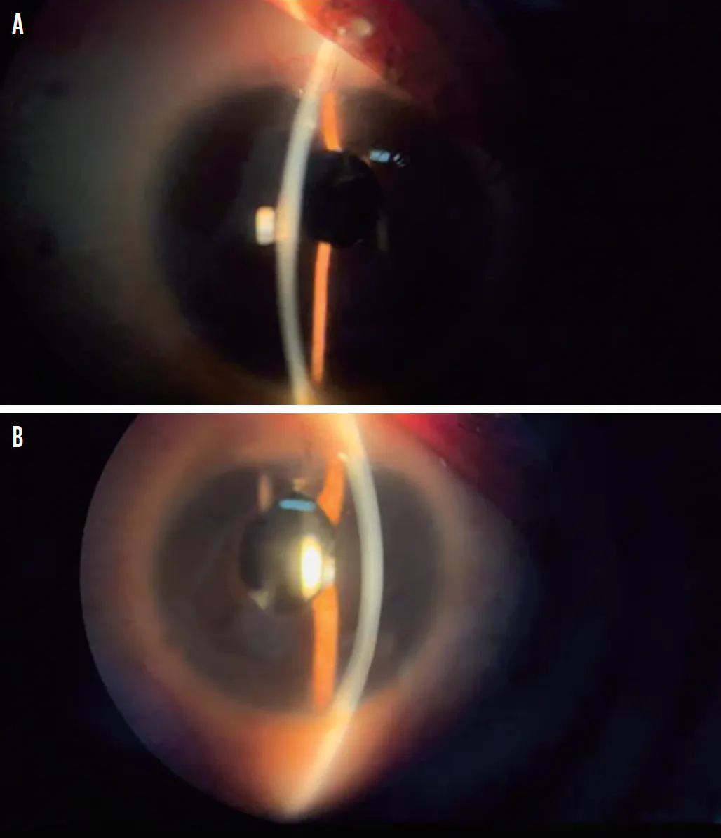
Figure 2. Postoperative anterior segment photographs of the right eye at month 14 (A) and the left eye at month 13 (B). Note the well-centered IOL, clear cornea, and absence of wound- or fixation-related complications in each eye.
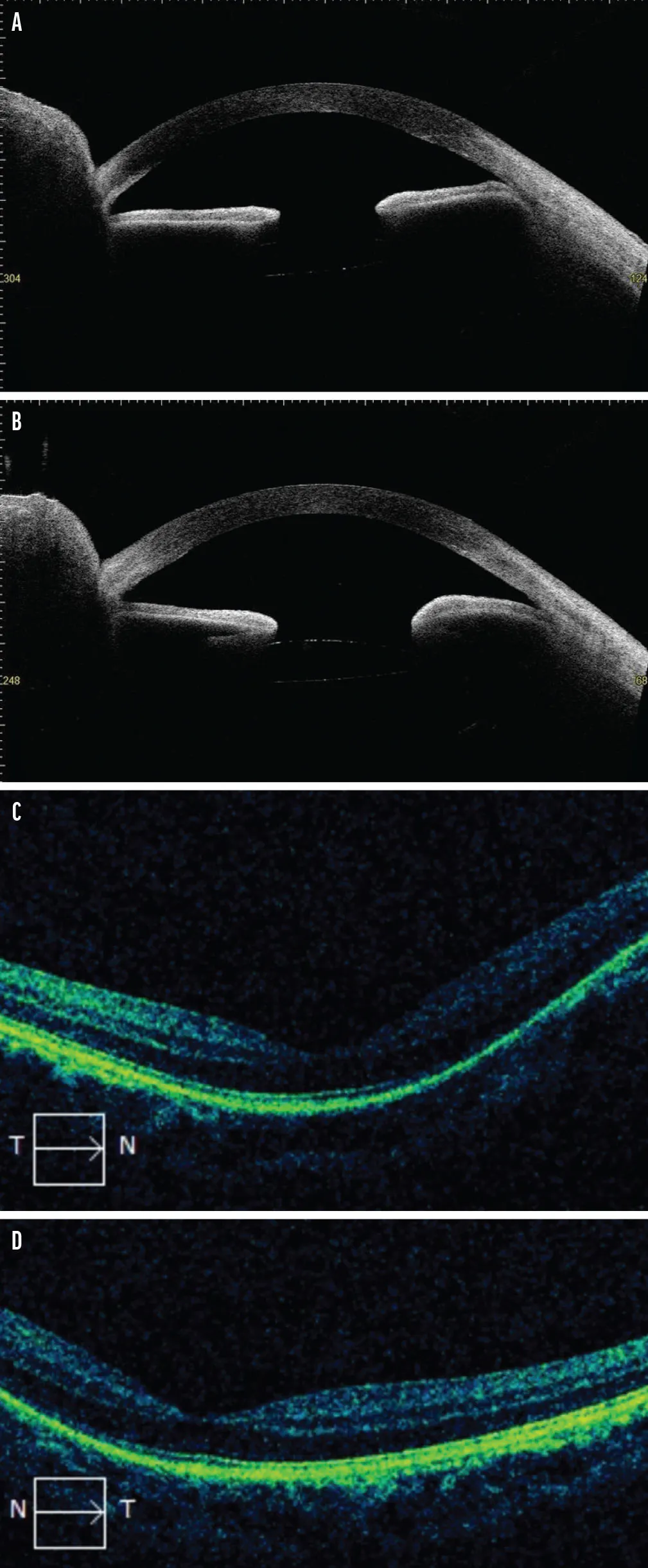
Figure 3. Anterior segment OCT shows the location of the IOL in the right eye at postoperative month 14 (A) and in the left eye at month 13 (B). Macula OCT of the right (C) and left eye (D). Note the stable intrascleral position of the IOL without tilt or decentration (A and B), and the preserved foveal contour without macular edema or structural abnormality (C and D).
Furthermore, we assessed macular thickness using spectral-domain OCT with a 512 x 128 macular cube scan protocol. To analyze patient’s internal limiting membrane and retinal pigment epithelium, we measured the central subfield thickness (CST), cube volume, and cube average thickness. The results for her right eye were 254 μm, 7 mm3, and 196 μm, respectively, whereas her left eye showed results of 258 μm, 10 mm3, and 279 μm, respectively. Although the CST of each eye was within the normal range, we observed notable asymmetry in the overall macular volume and average thickness, specifically a relative thinning of the macula in the right eye. This indicates that there was no complication of macular thickness after the implantation of the IOL.
SURGICAL CONSIDERATIONS FOR MICROCORNEA
Microcornea is defined as a corneal horizontal diameter less than 9 mm at birth, 10 mm at 2 years of age or older, or a vertical diameter less than 11 mm.4 This condition is often associated with zonular dialysis and subluxated cataracts, which may prevent the implantation of the IOL in the capsular bag.1 A sutured scleral-fixated IOL, a conventional alternative, may present a significant risk in eyes with microcornea, particularly with preexisting retinal conditions.1 Moreover, aphakic rehabilitation in eyes with microcornea can be challenging due to patient intolerance and poor contact lens fit, which is further worsened in patients with nystagmus. Therefore, secondary IOL implantation is often the more viable surgical alternative.5
The limited anterior chamber space in microcornea complicates traditional surgical techniques, particularly during haptic manipulation and IOL positioning. The reduced corneal diameter limits the space available for incision placement and complicates wound construction, requiring a shorter, more peripheral corneal tunnel and making instrument manipulation within the shallow anterior chamber more difficult, which may increase the risk of postoperative complications such as corneal edema and wound instability.6
Haptic trimming in our modified Yamane approach is a crucial technical modification that addresses these surgical challenges. A meta-analysis by Zhang et al compared the clinical outcomes of sutured scleral fixation with those of the Yamane technique for secondary IOL implantation and showed significantly improved BCVA at 1 month postoperative in the Yamane group in single-surgeon studies (P = .002).7 Beyond the inherent advantages of the Yamane technique, trimming the haptics provides additional benefits. A comparative study by Lin et al evaluating trimmed versus untrimmed haptics in the Yamane technique showed superior long-term IOL stability with trimmed haptics.8 In their study of 39 eyes, untrimmed haptics were frequently overlong, leading to haptic extrusion in three eyes (16%) in the untrimmed group versus none in the trimmed group at 24 months.
These results align with those of Kurimori et al, who reported two cases of severe IOL tilting following the Yamane technique and proposed that symmetrical shortening of the haptic edges to their most lateral extension point may reduce the tilt.9 This approach also improves refractive outcomes by reducing astigmatic power and achieving the planned refractive target. A prospective case series by Kumar et al further supported these findings, showing that custom haptic trimming with 6 mm optic foldable IOLs were safe to implant in microcornea eyes.1
Although glued scleral fixation provides benefits such as excellent flap closure, IOL centration, and stability without suture-related complications (particularly in cases with deficient or absent posterior capsules),10 our modified Yamane approach with haptic trimming offers adequate IOL stability, and the flanged haptic design ensures secure fixation within the scleral tunnels.
A USEFUL TECHNIQUE FOR OVERCOMING ANATOMIC CHALLENGES
Our patient’s condition of bilateral aphakia with nystagmus and microcornea was the result of bilateral cataract extraction during her childhood to prevent amblyopia. The extractions were the only procedure performed, likely because she lived in a rural area with limited access to specialized ophthalmic care. Despite her anatomic constraints, our use of the modified Yamane technique with haptic trimming without glued scleral fixation resulted in good visual outcomes and a significant improvement in her quality of life.
Acknowledgements: Institutional approval was not required for this case report. The authors obtained written informed consent from the patient for publication of this case for educational and research purposes.
1. Kumar DA, Agarwal A, Sivangnanam S, Chandrasekar R, Agarwal A. Implantation of glued intraocular lenses in eyes with microcornea. J Cataract Refract Surg. 2015;41(2):327-333.
2. Sinskey RM, Amin P, Stoppel J. Intraocular lens implantation in microphthalmic patients. J Cataract Refract Surg. 1992;18(5):480-484.
3. Brannan SO, Kyle G. Bilateral microcornea and unilateral macrophthalmia resulting in incorrect intraocular lens selection. J Cataract Refract Surg. 1999;25(7):1016-1018.
4. Robert MC, Colby K. Congenital anomalies. In: Colby K, ed. Corneal Diseases in Children. Springer International Publishing. 2017:69-85.
5. Alwohaibi NN, Aljindan MY, AlRashidi FN. Scleral fixated intraocular lens in aphakic patient with bilateral microcornea and microphthalmia. Int Med Case Rep J. 2021;14:365-369.
6. Khokhar S, Gupta S, Tewari R, et al. Scleral tunnel phacoemulsification: approach for eyes with severe microcornea. Indian J Ophthalmol. 2016;64(4):320-322.
7. Zhang C, Palka C, Zhu D, et al. Clinical outcomes in scleral fixation secondary intraocular lens with Yamane versus suture techniques: a systematic review and meta-analysis. J Clin Med. 2024;13(11):3071.
8. Lin H, Ye X, Huang X, et al. Long-term stability of intraocular lens with trimmed or untrimmed haptics in Yamane sutureless intrascleral fixation technique. Med Sci Monit. 2021 Mar 11;27:e928868.
9. Kurimori HY, Inoue M, Hirakata A. Adjustments of haptics length for tilted intraocular lens after intrascleral fixation. Am J Ophthalmol Case Rep. 2018;10:180-184.
10. Agarwal A, Kumar DA, Jacob S, Baid C, Agarwal A, Srinivasan S. Fibrin glue-assisted sutureless posterior chamber intraocular lens implantation in eyes with deficient posterior capsules. J Cataract Refract Surg. 2008;34(9):1433-1438.


_1784132761.jpg?auto=compress,format&w=70)


