


KEY TAKEAWAYS
- The 2026 Duke Fellows Advanced Vitreous Surgery (fAVS) Course provided 2 days of high-yield education, hands-on training, and collaborative discussion.
- The 2025 Robert Machemer/IRRF Fellowship awardee, Taariq Mohammed, MD, lectured on imaging of the vitreous using brillouin spectroscopy.
- The hands-on wet lab offered fellows the opportunity to refine surgical skills and gain exposure to emerging technologies, including 3D heads-up displays and new implants, and unique surgical techniques.
The 2026 Duke Fellows Advanced Vitreous Surgery (fAVS) Course, held March 27-28, 2026, in Durham, North Carolina, brought together leaders in vitreoretinal surgery, medical retina, uveitis, inherited retinal disease (IRD), and ocular oncology for high-yield education, hands-on training, and collaborative discussion. The program was directed by Lejla Vajzovic, MD, with co-directors Dilraj Grewal, MD; Xi Chen, MD, PhD; and Durga S. Borkar, MD, MMCi. The course brought together expert faculty and fellows for a dynamic educational experience featuring didactic lectures, interactive panel discussions, fellows’ surgical video presentations, and an extensive wet lab showcasing the latest innovations and technologies in vitreoretinal surgery.
MEDICAL RETINA EDUCATION
The meeting opened with a retinal pharmacotherapy session on advances in AMD, diabetic retinopathy, retinal vein occlusion, IRD, and uveitis. Margaret A. Chang, MD, MS, discussed tips for implanting the port delivery system with ranibizumab (Susvimo, Genentech/Roche). Mathew W. MacCumber, MD, PhD, highlighted the need for individualized care based on the DRCR Retina Network’s recommendations. Nieraj Jain, MD, then reviewed optogenetics, gene editing, and AI-enabled low vision technologies in IRD.
The next session focused on pediatric retina and ocular oncology. Safa Rahmani, MD, stressed that success in pediatric retina often depends on rapport, dilation, and flexibility. When possible, try to make the examination a game with your youngest patients, she advised. Basil K. Williams Jr, MD, gave a case-based talk on ocular tumors, emphasizing the importance of obtaining a history and thinking carefully before taking patients to the OR who have unexplained or unusual findings. Michael I. Seider, MD, noted that recognizing pseudomelanomas can be equally as important as identifying melanoma itself; careful assessment can prevent both overdiagnosis and unnecessary treatment.
The Robert Machemer, MD/International Retinal Research Foundation (IRRF) Fellowship was introduced by Cynthia A. Toth, MD. In the spirit of Dr. Machemer’s belief that “progress comes from doing the unconventional,” the fellowship supports surgical fellows in their transition to independent research early in their careers. The 2025 Robert Machemer/IRRF Fellowship awardee, Taariq Mohammed, MD, shared pearls on investing in strong mentorship relationships, developing research plans, and finding the right niche in academic ophthalmology (Figure). He then discussed his own research endeavors, which have pioneered imaging of the vitreous using brillouin spectroscopy.
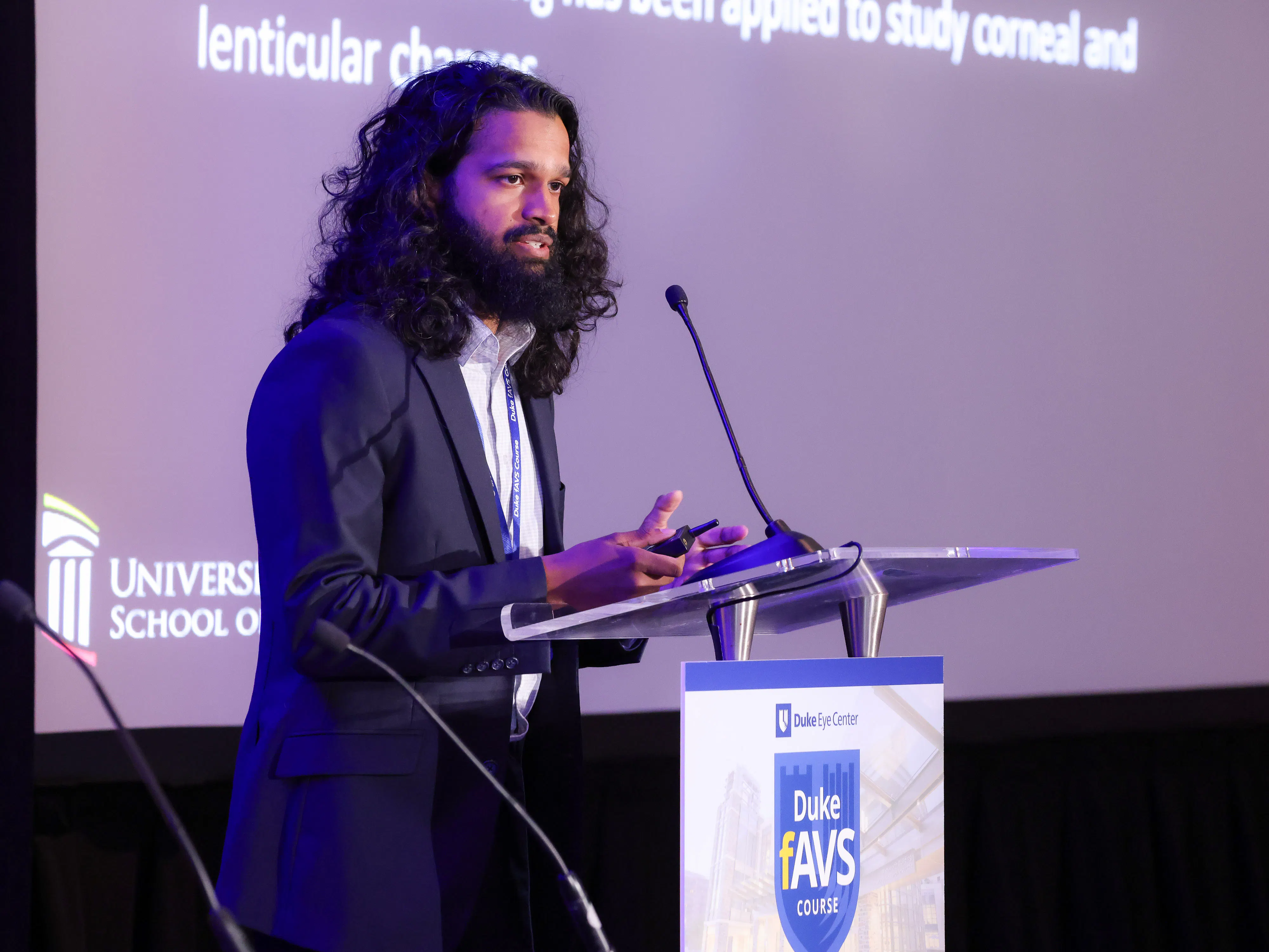
Figure. The 2025 Robert Machemer/IRRF Fellowship awardee, Dr. Mohammed, gave a talk on the biomechanics of the vitreous in vivo with the use of brillouin spectroscopy. Image courtesy of Kevin Caldwell Photography.
SURGICAL EDUCATION
The surgical curriculum emphasized practical approaches to complex conditions and OR issues such as ergonomics (by Dr. Williams), patients without a posterior vitreous detachment (by Dr. Chen), the costs of retinal detachment (RD) repair (by Jason Fan, MD, PhD), risk factors for proliferative vitreoretinopathy (by Dr. MacCumber), and core principles of surgery for diabetic tractional RD (by Yannek I. Leiderman, MD, PhD). Alice Zhang, MD, took to the podium to review her own surgical tips for managing lens subluxation and reviewed approaches to scleral buckle surgeries.
The next session focused on the practical aspects of building a career in retina, including coding and billing (by Dr. Chang), collaboration between academia and industry (by Dr. Toth), and the transition from fellowship to independent practice (by Dr. Rahmani).
The day concluded with the fellows’ surgical video session. Featured cases included combined tractional and rhegmatogenous RDs (by Mitchell Allphin, MD), submacular hemorrhage (by Beatriz Deud, MD), complex tractional RD with subretinal fibrotic bands and retained subretinal perfluorocarbon/oil (by Irmak Karaca, MD), biopsies (by Joseph Luvisi, MD), and an atypical intraoperative event in which subretinal fluid disappeared following perfluoro-octane injection due to a scleral rupture (by Linus Shen, MD).
MORE MEDICAL RETINA UPDATES
Day two began with discussions on treating wet AMD by Prithu S. Mettu, MD, and real-world data on pegcetacoplan (Syfovre, Apellis/Biogen) and avacincaptad pegol (Izervay, Astellas) by Eleonora G. Lad, MD, PhD. Majda Hadziahmetovic, MD, explored the role of AI in diabetic retinopathy screening, noting that it can identify changes before symptoms develop. She then highlighted the Duke Remote Retinal Imaging Program and other initiatives. Amol A. Sura, MD, presented a stepladder approach to immunotherapy in uveitis based on the ADVISE trial, and Oleg Alekseev, MD, PhD, reviewed cases in which genetic testing for IRD influenced patient counseling and management.
In the next session, Ramiro S. Maldonado, MD, gave an overview of the evolving strategies and current ongoing trials for treating Stargardt disease, including oral therapies, cell replacement, optogenetics, and the PRIMA implant (Science Corporation). Miguel A. Materin, MD, led a case-based discussion on differentiating benign versus malignant lesions in the eye. The session concluded with a case panel, including a discussion of retina pigment epithelium tears after intravitreal injections, building a list of differentials for hereditary maculopathies, recognizing salient features of toxic maculopathies (particularly from pentosan polysulfate), and managing delayed onset endophthalmitis after cataract surgery.
WET LAB AND EMERGING TECHNOLOGIES
The wet lab experience, a cornerstone of the course, offered fellows the opportunity to refine surgical skills and explore emerging technologies. Stations included subretinal delivery, membrane peeling, scleral fixation techniques, port delivery system implantation, and various intravitreal and suprachoroidal implants/injections. Attendees also engaged with next-generation surgical systems such as the Bausch + Lomb’s SeeLuma 3D heads-up digital visualization platform, Alcon’s Unity Vitreoretinal Cataract System, DORC’s EVA Nexus machine, and Zeiss’ Rescan iOCT and Artevo 800.
The course concluded with the well-loved annual, “Great Job Search Panel,” moderated by Sharon Fekrat, MD, FASRS, during which panelists shared their pearls for job contract negotiations and starting a position as a new attending. The experts explored various practice setting considerations, the importance of clinic schedules, and how to find research opportunities. They all agreed that who you work with is the most important factor when searching for the right job.
SAVE THE DATE
13th annual Fellows Advanced Vitreous Surgery (AVS) Course and 24th Duke AVS Course
March 18-20, 2027
_1784132761.jpg?auto=compress,format&w=70)


