





KEY TAKEAWAYS
- Spontaneous lens absorption is a rare complication that can occur in association with various conditions, including hypermature or traumatic cataract, as well as in the late stages of uveitis or other infectious diseases.
- Spontaneous lens absorption is considered unlikely in the presence of an intact lens capsule and is rather facilitated by its rupture, whether macroscopic or microscopic, thus allowing the leakage of lens material.
- In each case the authors report, a desiccation cataract was present during the postoperative period following 23-gauge pars plana vitrectomy for retinal detachment repair, followed by a progressive densification of the cataract and, later, spontaneous transition to an aphakic state.
Spontaneous lens absorption is a rare complication that can occur in association with various conditions, including hypermature or traumatic cataract, as well as in the late stages of uveitis or other infectious diseases.1-3 Lens absorption following pars plana vitrectomy (PPV) is an unusual finding that has rarely been reported in the literature, with one case describing spontaneous cataract absorption diagnosed 7 years after the initial vitrectomy and silicone oil tamponade.4
We report here two similar cases of spontaneous lens absorption following PPV with gas tamponade to repair rhegmatogenous retinal detachment, which were subsequently treated with IOL implantation.
CLINICAL FEATURES
Case No. 1
A 54-year-old man with a history of systemic hypertension presented to our clinic with a sudden decrease in vision in his left eye. His VA was 20/25 OD and counting fingers at 1 m OS at presentation. On examination, the anterior segment was unremarkable with a transparent crystalline lens in each eye. Fundus photography revealed a superior macula-off retinal detachment in his left eye, extending from the 10 to 2 clock hours with multiple peripheral tears; the right fundus was normal.
The patient underwent 23-gauge transconjunctival PPV with C2F6 gas tamponade. No lens manipulation occurred during the surgery. On postoperative day 1, his VA was counting fingers at near OS, with mild corneal edema, a desiccation cataract and gas filling the entire vitreous cavity. At the follow-up appointment 1 week later, his VA improved to counting fingers at 3 m OS; the cornea was clear, and there was still a desiccation cataract, gas filling four-fifths of the vitreous cavity, and a flat retina. However, at the next visit 1 month postoperatively, VA declined to counting fingers at 1 m OS; on examination, the cornea was clear with a progression of the cataract, residual gas in the vitreous cavity, and a flat retina. At the 3-month follow-up, VA had further decreased to hand motion OS. The cornea remained clear, but the cataract had become total, obscuring the fundus. Ocular ultrasound showed a flat retina.
The patient was scheduled for cataract surgery in his left eye but was lost to follow up (LTFU). However, he was found and reexamined more than a year later, at which point his VA had spontaneously improved from hand motion to counting fingers at 1 m OS. His refractive error was not measurable, as the visual axis was obstructed by residual capsular opacities. The anterior segment showed a clear cornea, a deepened and quiet anterior chamber, iridodonesis, synechiae at the 4 and 5 clock hours, and aphakia with a ripped posterior lens capsule (Figure 1). The retina was flat. No trace of the lens was noted either on the ophthalmoscopic examination or echographically, and a thorough questioning ruled out any intervention during the period of LTFU.
The patient’s aphakia was corrected via IOL implantation in the sulcus and surgical clearing of the visual axis with good functional vision recovery.
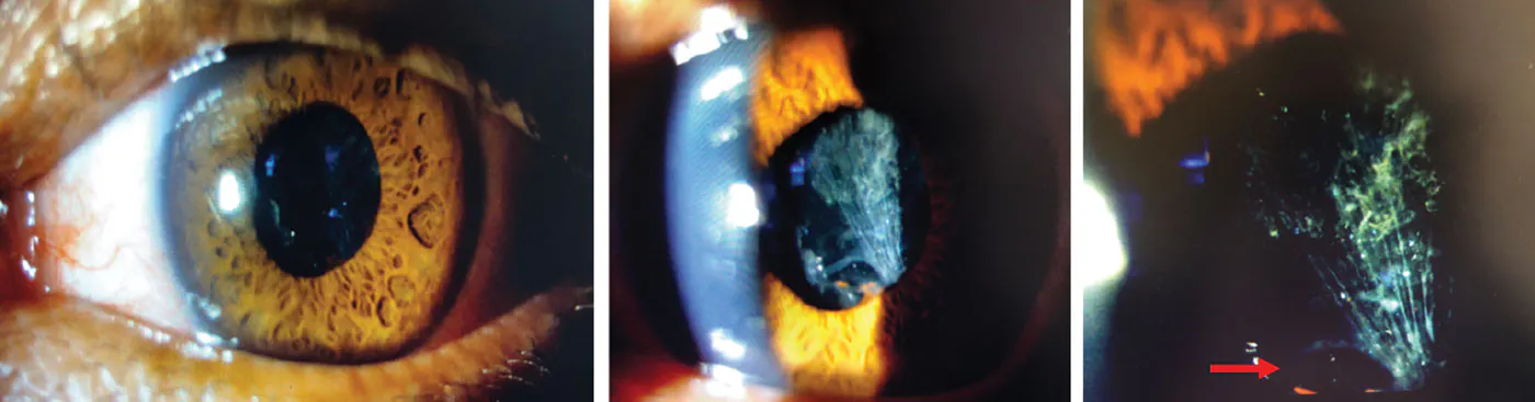
Figure 1. Anterior segment examination of the first patient’s left eye shows an empty capsular bag with opacification of the capsular remnant. Note the small breach in the inferonasal region (arrow).
Case No. 2
A 49-year-old woman with an amblyopic left eye, likely due to a traum during childhood, presented with a retinal detachment in her better-seeing right eye. Her BCVA was 20/40 OD and counting fingers at 2 m OS. The crystalline lens was transparent in each eye. Her right eye showed a macula-on retinal detachment, extending from the 2 to 7 clock hours with multiple peripheral retinal tears. Fundus examination of her left eye was normal.
The patient underwent 23-gauge transconjunctival PPV with C2F6 gas tamponade. No lens manipulation occurred during the surgery. On postoperative day 1, her VA was counting fingers at 0.3 m OD, with mild corneal edema and a desiccation cataract, a complete gas fill, and a flat retina. At the 1-month follow-up, her VA was counting fingers at 0.5 m OD, with a clear cornea, a denser desiccation cataract, residual gas in the vitreous cavity, and a flat retina. At the 2-month follow-up, her VA declined to hand motion OD. The cornea remained clear, but the cataract had become total, with no fundus visibility. The ocular echography showed a flat retina. The ocular findings remained stable during the next two follow-up visits at 5 and 7 months postoperative.
The patient was scheduled for cataract surgery for her right eye but was LTFU. She was found and reexamined about 1 year later, at which time her uncorrected VA improved from hand motion OD to counting fingers at 0.5 m OD. Her BCVA after correction of the measured refractive error of +9.75 (-2 at 18°) was 20/63 OD. The anterior segment examination revealed a clear cornea, a deepened and quiet anterior chamber, iridodonesis, and aphakia associated with a large tear in the posterior capsule of the lens (Figure 2). The retina was flat. No trace of the lens was found either on the ophthalmoscopic examination or echographically. A thorough patient history ruled out any eye surgery or manipulation during the period of LTFU. The patient’s aphakia was corrected by IOL implantation in the sulcus with good functional vision recovery.
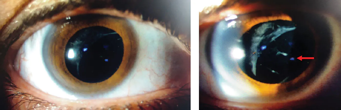
Figure 2. Anterior segment examination of the second patient’s right eye shows an empty capsular bag with a large posterior lens capsule tear (arrow).
PROPOSED MECHANISMS OF LENS ABSORPTION
In each of the cases reported here, a desiccation cataract was present during the postoperative period following 23-gauge PPV with gas tamponade. The cataract became progressively denser, and VA decreased to hand motion within 3 months for each patient. However, VA improved without any external intervention to counting fingers at 1 m for the first patient and counting fingers at 0.5 m for the second, which was corrected to 20/63. This improvement coincided with the transition from the cataractous lens state to the aphakic state, observed approximately 1 year after the initial surgical intervention, as both patients had a period of LTFU. This led us to conclude that spontaneous absorption of the cataractous lens occurred following PPV, as no external intervention occurred during this period.
The mechanism underlying spontaneous lens absorption has not been fully elucidated, but it has been theorized that structural, chemical/osmotic, and immunological events likely facilitate this process.2
Spontaneous lens absorption is thought to be unlikely in the presence of an intact lens capsule; conversely, absorption may be facilitated following damage to the lens capsule as in traumatic cases and late, hypermature cataract.2 In systemic infectious cases, such as leptospirosis and rubella, it is thought to be a response to either the bacteria or antibodies.5,6 These pathophysiological mechanisms could allow for a macroscopic or microscopic leakage of crystalline lens material through a breached or abnormally permeable lens capsule, thus leading to its absorption.
The most likely explanation in our patients who underwent PPV is a breach in the posterior lens capsule that went unnoticed during surgery, thus facilitating lens absorption and bringing these cases closer to post-traumatic ones. This is further corroborated by the evidence of a tear in the posterior lens capsule in both patients observed after the period of LTFU.
CAREFULLY RULE OUT OTHER POSSIBILITIES
As lens damage is not uncommon during PPV in phakic patients, these cases draw attention to the possibility of spontaneous lens absorption in the postoperative period following PPV.
1. Painter SL, Imrie FR, Mayer EJ. Lens reabsorption following self-induced needling and subsequent intracapsular secondary intraocular lens placement. J Cataract Refract Surg. 2008;34(5):868‑870.
2. Marlow SB. Spontaneous absorption of cataract. Trans Am Ophthalmol Soc. 1952;50:283‑293.
3. Boger WP, Petersen RA, Robb RM. Spontaneous absorption of the lens in the congenital rubella syndrome. Arch Ophthalmol Chic Ill 1960. 1981;99(3):433‑434.
4. Liu L, Wang F, Gu XY. A case of spontaneous lens absorption following cataract complication after vitrectomy combined with silicone oil tamponade. Zhonghua Yan Ke Za Zhi. 2025;61:911–913.
5. Smith GT, Shun-Shin GA, Bron AJ. Spontaneous reabsorption of a rubella cataract. Br J Ophthalmol. 1990;74(9):564‑565.
6. Rathinam S, Namperumalsamy P, Cunningham E. Spontaneous cataract absorption in patients with leptospiral uveitis. Br J Ophthalmol. 2000;84(10):1135‑1141.

_1784132761.jpg?auto=compress,format&w=70)


