




KEY TAKEAWAYS
- Children with inherited retinal diseases (IRDs) tend to experience faster disease progression and significant diagnostic delays due to a mismatch between patient/caregiver complaints and clinical examination.
- Handheld imaging, such as swept-source OCT and portable full-field electroretinography, are under investigation as tools to improve diagnosis and disease monitoring in the pediatric IRD population.
- The field needs additional studies to validate functional endpoints that are feasible in a pediatric population, or structural endpoints that are more objective and do not rely on patient cooperation.
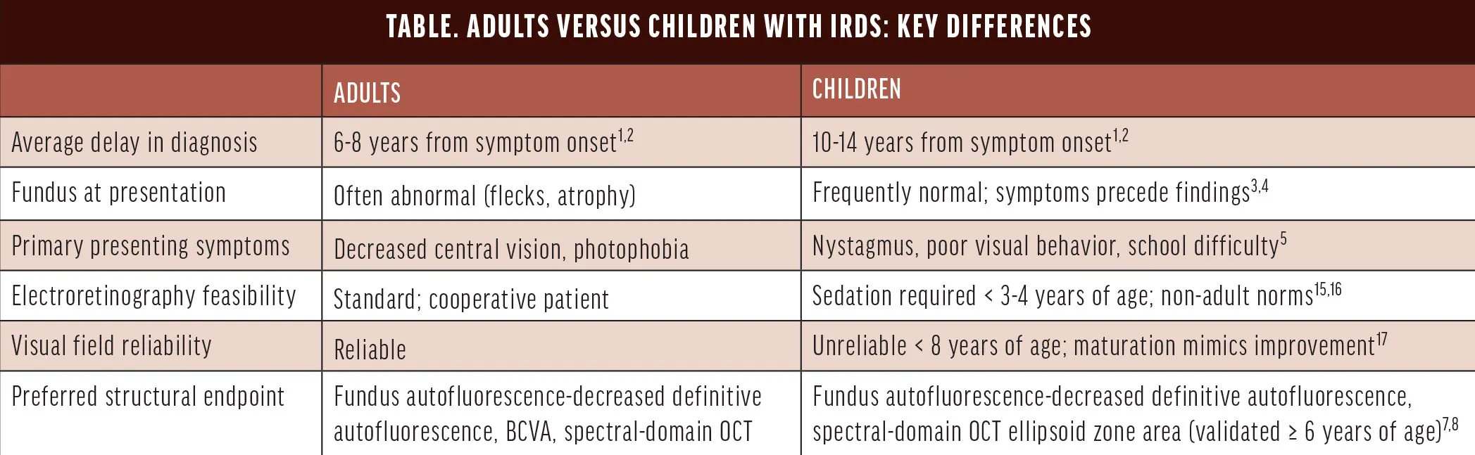
The standard diagnostic and management framework for adults with inherited retinal diseases (IRDs) is well established, with symptom-driven referral, multimodal imaging, genetic testing, and, for a small subset, enrollment in clinical trials. However, children with an IRD represent a different clinical challenge, with distinct diagnostic pathways, phenotypic expression, and barriers to trial access (Table). A key limitation is the lack of imaging and functional technologies for children, making early and reliable disease characterization difficult. Here, we examine the differences in diagnostic delays, phenotypes, and management in children with IRDs compared with adults.
DIAGNOSIS AND CLINICAL PRESENTATION
Children with IRDs face significant diagnostic delays. For example, in our study of 87 patients with molecularly confirmed Stargardt disease at Duke University, the mean time from symptom onset to first IRD specialist evaluation was 9 years for early-onset patients (presenting in early childhood) and 14.4 years for intermediate-onset patients (presenting in adolescence) compared with 6.4 years for late-onset patients (presenting after age 45). The mean time from symptom onset to molecular diagnosis was even longer at 10.9 and 14 years for early- and intermediate-onset, respectively, compared with 6.7 years for late-onset.1 In another study of 1,096 adult patients with an IRD across the United Kingdom and Latin America, the mean time from symptom onset to molecular diagnosis was 6.4 ± 9.1 years, with 57% waiting more than 1 year. Notably, children were 2.1 times more likely to wait more than 1 year for a diagnosis compared with adults.2
The reasons for this delay are multifactorial. Young children cannot articulate visual symptoms, and the presenting complaint is often parental concern about a child’s visual behavior, not a self-report of central scotoma or photophobia, as is more typical for adults. In children, nystagmus (76%) and abnormal visual behavior (28%) often lead to initial evaluation by a pediatric ophthalmologist and pediatric neurologist rather than a retina specialist.3
In addition, the nonspecific visual symptoms can be accompanied by a normal fundus examination, making diagnosis based on clinical examination more challenging. Early-onset Stargardt disease frequently presents with vision loss and cone-rod dysfunction on electroretinogram (ERG) that precedes any visible fundus abnormality.4,5 In a series of 31 patients with early-onset Stargardt disease without fundus findings, 87% experienced delayed diagnosis (median 3 years), and 94% had visited more than two hospitals misdiagnosed as amblyopia, functional vision loss, or optic neuropathy.4 Even in Leber congenital amaurosis, profound vision loss, nystagmus, and an undetectable ERG appear in the first 6 months of life, yet the fundus can appear normal at initial examination.3
Meanwhile, intermediate- and late-onset Stargardt disease show characteristic fundus autofluorescence (FAF) changes, while retinitis pigmentosa in adults shows ellipsoid zone (EZ) loss correlating with night blindness and peripheral visual field constriction.6,7 Patients with late-onset Stargardt disease were misdiagnosed with AMD in 22% of cases, but the clinical presentation (macular atrophy, presence of flecks) tends to be interpretable enough to eventually prompt appropriate workup.1
Furthermore, IRD progression can be faster in children compared with adults. For instance, annual EZ area loss in childhood-onset Stargardt disease averaged 1.20 ± .29 mm²/year, approximately three times the adult progression rate, and FAF showed that decreased definitive autofluorescence lesion growth in childhood-onset patients progresses roughly twice as fast as in adult-onset cohorts.8-10 In our study, nearly 90% of early-onset patients lost subfoveal EZ integrity before reaching the IRD clinic, compared with 30% of adults with late-onset Stargardt disease.1
THE ROLE OF IMAGING
Because most diagnostic tests are optimized for adults and difficult to perform in children, OCT imaging has become crucial to detect photoreceptor degenerative changes across different genotypes. Currently, there is only one commercially available handheld spectral-domain OCT (Envisu C2300, Leica), which was approved by the FDA in 2007.11 However, its acquisition speed is suboptimal for children with IRDs, particularly those with nystagmus. To address this limitation, Duke has developed an investigational noncontact, 1,064 nm wavelength, high speed (400 kHz) handheld swept-source OCT (SS-OCT). Our group evaluated the feasibility of the Duke handheld SS-OCT and showed that successful imaging was achieved in 81.1% of awake children with IRDs, providing a unique opportunity to evaluate early retinal changes without the need for examination under anesthesia.12 Other investigational ultra-widefield handheld SS-OCTs (eg, Theia T1-W, Theia Imaging) are currently being developed and have been shown to successfully image infants and children, both in clinic and under anesthesia.13
To bridge the gap between the International Society for Clinical Electrophysiology of Vision (ISCEV) standard protocol and the Great Ormond Street Hospital (GOSH) approach, we are integrating portable full-field ERG into our pediatric retinal evaluation. ISCEV-ERG remains the standard but requires prolonged dark adaptation, corneal electrodes, and sustained cooperation, often necessitating examination under anesthesia in young children. In contrast, the GOSH-ERG enables rapid, awake testing using skin electrodes, minimal dark adaptation, and handheld stimulation, while maintaining strong concordance with ISCEV responses (Figure). This approach reduces reliance on anesthesia and expands access to functional retinal assessment in pediatric IRD.
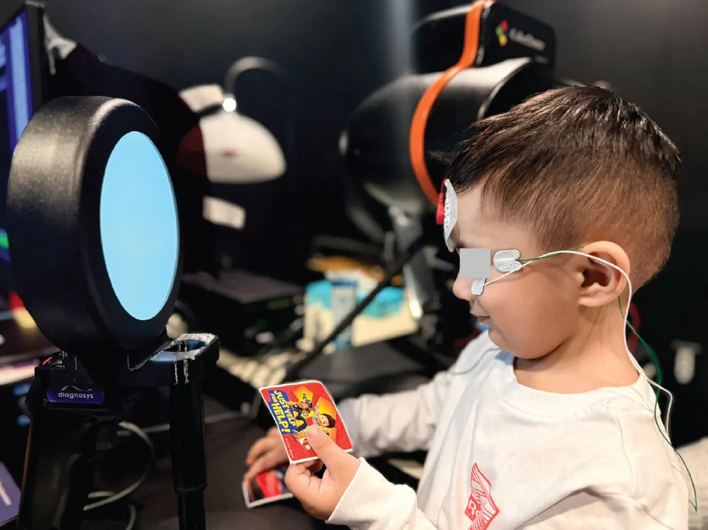
Figure. Portable full-field ERG performed in a pediatric patient using periocular skin electrodes and handheld flash stimulation, demonstrating a child-friendly, non-mydriatic approach with minimal dark adaptation. Image courtesy of RML lab (Retina Machine Laboratory).
GENETIC TESTING AND COUNSELING
Genetic testing is key to diagnosing IRD, but access remains limited, particularly for children. While commercial IRD gene panels (200 to 350 genes) and sponsored programs have expanded availability, children are less likely to go undergo testing due to referral gaps and logistical barriers. In a nationwide survey of pediatric retina and IRD specialists, children on Medicaid were perceived to have greater difficulty obtaining genetic testing despite state-level analysis indicating coverage in approximately two-thirds of states (68.6%); most providers were uncertain about this coverage, highlighting variability and poor transparency across policies. Cost concerns exceeded uncertainty in result interpretation or management as well.14
CLINICAL TRIALS AND MANAGEMENT
Gene therapies, antisense oligonucleotides, and small molecules are in active clinical trials for Stargardt disease, X-linked retinitis pigmentosa, Usher syndrome 2A, and CNGB3/CNGA3 achromatopsia. Most trials were designed for adults: Eligibility typically requires BCVA between approximately 20/32 and 20/400 in the study eye, cooperative full-field ERG and Goldmann visual field testing, and a minimum of 18 years of age. For adults who reach an IRD specialist with moderate vision loss and preserved photoreceptor structure, these windows are generally accessible.
Children face a paradoxical exclusion from clinical trials in Stargardt disease: Their disease progression is often too advanced based on OCT imaging but too early based on FAF. In our Stargardt cohort, nearly 90% of early-onset patients had lost subfoveal EZ integrity at presentation, while FAF frequently showed questionable decreased autofluorescence.1 Because many Stargardt trials require a minimum area of decreased definitive autofluorescence, these children do not meet enrollment criteria despite established photoreceptor damage.2 Functional tests are also more challenging to perform on children. ERG requires sedation in young children and produces results not directly comparable with adult normative data (ie, smaller amplitudes, delayed implicit times).15,16 Goldmann visual field testing is similarly unreliable before 7 to 8 years of age.17 Currently, full-field stimulus testing requiring no fixation is the validated endpoint for severe pediatric vision loss. It was accepted as a clinical trial endpoint by the FDA for voretigene neparvovec (Luxturna, Spark) and is feasible in school-age children; the multi-luminance mobility test is feasible from 4 years of age.18-21 The use of EZ width measurements remains exploratory. Overall, this highlights the need for additional studies to validate other functional endpoints that are feasible in a pediatric population, or ideally structural endpoints that are more objective and do not rely on patient cooperation.
TAKE THE PICTURE
Children with IRDs face unique challenges, such as diagnostic delays, potential faster IRD progression, and difficulty with functional testing for trial endpoints. Some solutions for addressing these challenges include educating all pediatric eye providers on the importance of obtaining multimodal imaging, such as OCT and FAF, in children with visual impairment, even in the presence of a normal fundus examination. This can streamline referrals to IRD specialists and genetic testing, and further development of structural endpoints for clinical trials in IRDs.
1. Li AS, Morales PC, Estrada-Puente C, Maldonado RS. Factors influencing the delayed diagnosis of Stargardt disease and impact on therapeutic opportunities. Retina. 2026;46:367-372.
2. Daich Varela M, Georgiou M, Hashem SA, et al. Diagnostic odyssey of more than 1000 patients with inherited retinal diseases. Ophthalmology. 2024;131(2):213-222.
3. Kumaran N, Moore AT, Weleber RG, Michaelides M. Leber congenital amaurosis/early-onset severe retinal dystrophy: clinical features, molecular genetics, and therapeutic interventions. Br J Ophthalmol. 2017;101(9):1147-1154.
4. Bax NM, Lambertus S, Cremers FPM, Klevering BJ, Hoyng CB. The absence of fundus abnormalities in Stargardt disease. Graefes Arch Clin Exp Ophthalmol. 2019;257:1147-1157.
5. Lambertus S, van Huet RAC, Bax NM, et al. Early-onset Stargardt disease: phenotypic and genotypic characteristics. Ophthalmology. 2015;122(2):335-344.
6. Pas JAAH, Dhooge PPA, Hoyng CB. Clinical classification of Stargardt disease. Graefes Arch Clin Exp Ophthalmol. 2024;262(5):1377-1379.
7. Sakai D, Takagi S, Hirami Y, Nakamura M, Kurimoto Y. Use of ellipsoid zone width for predicting visual prognosis after cataract surgery in patients with retinitis pigmentosa. Eye (Lond). 2023;37(1):42-47.
8. Tanna P, Georgiou M, Strauss RW, et al. Cross-sectional and longitudinal assessment of the ellipsoid zone in childhood-onset Stargardt disease. Transl Vis Sci Technol. 2019;8(2):1.
9. Georgiou M, Kane T, Tanna P, et al. Prospective cohort study of childhood-onset Stargardt disease: FAF imaging, progression, comparison with adult-onset disease, and disease symmetry. Am J Ophthalmol. 2020;211:159-175.
10. Daich Varela M, Laich Y, Hashem SA, et al. Prognostication in Stargardt disease using fundus autofluorescence. Ophthalmology. 2023;130(11):1182-1190.
11. Bioptigen receives FDA premarket approval for new OCT system [press release]. Healio. January 29, 2007. Accessed May 26, 2026. www.healio.com/news/ophthalmology/20120331/bioptigen-receives-fda-premarket-approval-for-new-oct-system
12. Estrada-Puente C, Morales PC, Narawane A, et al. Feasibility of ultracompact hand-held swept-source optical coherence tomography (HH-SS-OCT) for evaluating early onset retinal dystrophies (EORDs) in children. Invest Ophthalmol Vis Sci. 2025;66(8):1492.
13. Li, AS, Imperio R, Tran-Viet D, et al. Imaging infants and children with investigational handheld optical coherence tomography with widefield lens: a pilot study [published online ahead of print March 30, 2026]. Graefes Arch Clin Exp Ophthalmol.
14. Li AS, Maldonado RS, Lee J, Justin GA, Chen X, Toth CA, Vajzovic L. Barriers to genetic testing in children with inherited retinal diseases. Presented at: Ophthalmic Genetics Study Club; November 3, 2023.
15. McCulloch DL, Marmor MF, Brigell MG, et al. ISCEV standard for full-field clinical electroretinography (2015 update). Doc Ophthalmol. 2015;130(1):1-12.
16. Robson AG, Frishman LJ, Grigg J, et al. ISCEV standard for full-field clinical electroretinography (2022 update). Doc Ophthalmol. 2022;144(3):165-177.
17. Dedania VS, Liu JY, Schlegel D, et al. Reliability of kinetic visual field testing in children with mutation-proven retinal dystrophies: Implications for therapeutic clinical trials. Ophthalmic Genet. 2018;39(1):22-28.
18. Roman AJ, Cideciyan AV, Wu V, Garafalo AV, Jacobson SG. Full-field stimulus testing: role in the clinic and as an outcome measure in clinical trials of severe childhood retinal disease. Prog Retin Eye Res. 2022;87:101000.
19. Maguire AM, Russell S, Wellman JA, et al. Efficacy, safety, and durability of voretigene neparvovec-rzyl in RPE65-associated inherited retinal dystrophy: results of phase 1 and 3 trials. Ophthalmology. 2019;126(9):1273-1285.
20. Chung DC, McCague S, Yu ZF, et al. Novel mobility test to assess functional vision in patients with inherited retinal dystrophies. Clin Exp Ophthalmol. 2018;46(3):247-259.
21. Russell S, Bennett J, Wellman JA, et al. Efficacy and safety of voretigene neparvovec-rzyl in RPE65-mediated inherited retinal dystrophy: a randomised, controlled, open-label, phase 3 trial. Lancet. 2017;390:849-860.

_1784132761.jpg?auto=compress,format&w=70)


