




Key Takeaways
- When planning surgery for tractional retina detachments (TRDs), OCT imaging, ultrasound (when needed), possible cataract extraction, and anti-VEGF injection can help map out the extent of the traction, assess the status of the hyaloid, and regress active neovascularization.
- Useful tools in TRD surgery include a delaminating spatula, a high-magnification contact lens, MPC vertical scissors, a lighted pick, a Finesse Flex Loop (Alcon), and dextrose added into the infusion bottle.
- Tamponade selection should favor gas over silicone oil in most complex traction/rhegmatogenous RD cases, as long-acting gas provides 6 to 8 weeks of tamponade and avoids the need for oil removal surgery.
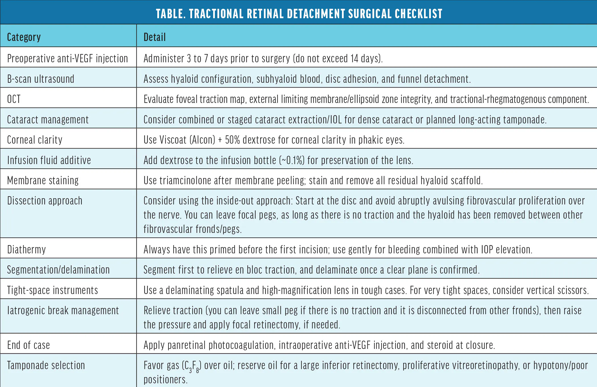
Diabetic tractional retinal detachments (TRDs) are some of the most technically demanding cases in vitreoretinal surgery. Each eye presents a unique constellation of posterior hyaloid configuration, vitreoretinal adhesions, fibrovascular proliferation, and neovascular activity. Success demands technical proficiency, systematic preoperative planning, thoughtful selection of instrumentation, and sound intraoperative decision making. In this article, we share our considerations during the key phases of complex diabetic vitrectomy (Table).1
PREOPERATIVE PLANNING
Preoperative OCT is invaluable for mapping the extent of the traction, evaluating the foveal contour (including the presence of an epiretinal membrane), identifying focal fibrovascular pegs overlying the macula (and an associated plane for dissection), and identifying any associated tractional-rhegmatogenous component. The integrity of the external limiting membrane and ellipsoid zone can also inform counseling on visual prognosis, which is often limited.
When media clarity is compromised by dense vitreous hemorrhage, B-scan ultrasonography is essential. Key features to assess using this tool include the configuration of the posterior hyaloid, the extent of the subhyaloid hemorrhage, the presence and location of a funnel detachment, and the relationship of fibrovascular tissue to the disc.
While practice patterns vary, preoperative bevacizumab (Avastin, Genentech/Roche) administered 3 to 7 days prior to surgery reduces intraoperative bleeding by regressing active neovascularization.2 Beyond 10 to 14 days, however, the risk of accelerating the contraction of fibrovascular membranes and worsening traction increases. Because these patients often carry significant comorbidities, we recommend confirming transportation, medical clearance, and scheduling requirements (eg, hemodialysis) prior to injection.
If a cataract is present, combined or staged phacoemulsification can be considered, depending on the patient’s age, the visual significance of the cataract, the quality of posterior segment visualization, and whether a longer-acting tamponade is planned. Removing a dense lens dramatically improves visualization, maneuverability, and effectiveness of peripheral vitreous base shaving, which can otherwise serve as a nidus for recurrent neovascularization postoperatively.
To ensure corneal clarity, consider using a dispersive viscoelastic applied to the corneal surface, along with 5 cc to 10 cc of topical 50% dextrose solution on the cornea (when available) under the viscoelastic layer to maintain corneal clarity and prevent corneal edema throughout the case.
ESSENTIAL INSTRUMENTATION
We favor 25-gauge instrumentation for most cases, balancing efficient vitreous removal with instrument stability. However, 27-gauge offers a unique advantage with an optimized cutter mouth-to-tip distance for segmentation of adherent membranes. In cases with dense vitreous hemorrhage or thick membranes requiring more aggressive segmentation, 23-gauge provides greater cutting efficiency and instrument rigidity. Know your system and have a plan for conversion.
The following additional instruments are essential to our complex diabetic TRD setup in the OR:
- A delaminating spatula is essential for developing safe planes, which is the cornerstone of success in TRDs.
- A high-magnification contact lens can optimize visualization of the posterior pole during membrane dissection in the macula, especially for plane recognition around retinal vessels, the optic nerve, fovea, and just outside the arcades to prevent breaks.
- MPC vertical scissors allow access to membranes tightly adherent to the arcade vessels and can be used closed for blunt dissection (Figure 1). Vertical scissors are preferred for segmentation perpendicular to the retinal surface in tight spaces that other instruments cannot access. Because they are handled by the foot pedal, they can be very precise. The edges are relatively sharp, so caution is needed to prevent iatrogenic breaks. These scissors can be used alongside the delaminating spatula to ensure a safe plane is created before delamination.
- In select cases, a lighted pick can illuminate the surgical field and function as a dissection instrument for developing tissue planes beneath the hyaloid. The sharp edge can create breaks in the fragile diabetic retina, so exercise extreme caution when using it.
- The Finesse Flex Loop (Alcon) is useful for peeling sticky residual hyaloid membranes, mobilizing preretinal blood clots resistant to aspiration, and atraumatic tissue mobilization in tight anatomical spaces.
- Dextrose in the infusion bottle creates a hyperosmolar environment that helps tamponade neovascular vessels and reduce intraoperative bleeding. Using BSS+ (Alcon) adds dextrose, bicarbonate, and glutathione to the concentrate to preserve corneal endothelial cells and the lens.
After membranes are peeled, intravitreal triamcinolone injected into the vitreous cavity provides invaluable staining of any residual hyaloid scaffold and cortical vitreous. This step is critical because residual vitreous scaffold leaves persistent traction and creates a nidus for fibroglial proliferation. It must be removed before gas or oil tamponade is placed.
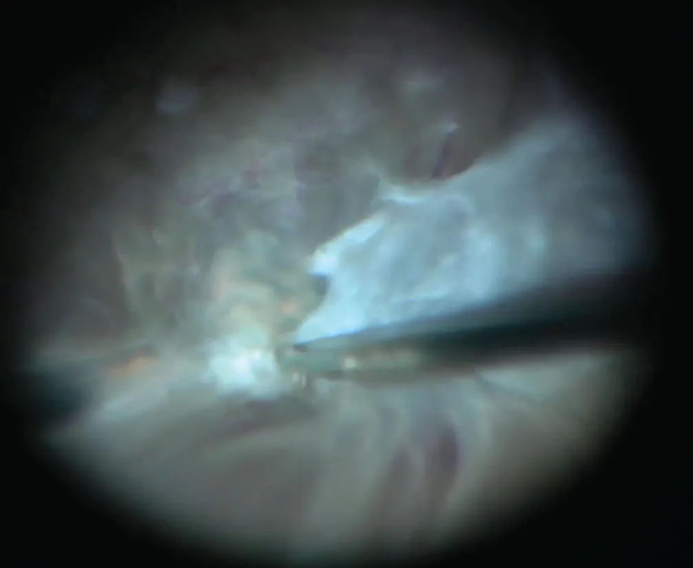
Figure 1. MPC scissors can be useful to access membranes that are tightly adherent to the arcade vessels and can aid in blunt dissection when closed.
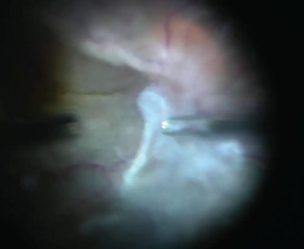
Figure 2. Surgeons can start posteriorly to elevate the fibrovascular tissue and create a plane for dissection.
SURGICAL APPROACH: INSIDE-OUT VERSUS OUTSIDE-IN
We favor the inside-out (posterior-to-anterior) approach for nearly all TRD cases. Starting at the posterior pole—particularly at the disc, where the hyaloid is often most firmly attached—allows the surgeon to develop a safe working plane under direct visualization before moving peripherally (Figure 2).
The critical principle is to never aggressively avulse fibrovascular proliferation over the optic nerve. The vascular supply to the disc is unforgiving. Instead, use internal limiting membrane forceps to apply gentle, tangential traction to remove the hyaloid/fibrovascular proliferation off the nerve, with diathermy immediately after. The moment you feel resistance at the disc, stop, consider using the soft-tip extrusion cannula on low vacuum, and be patient. Alternatively, stiffen the tissue with viscoelastic, apply diathermy to vessels under tension (while carefully avoiding the nerve itself), and then proceed.
Segmentation versus delamination is a key intraoperative decision. In general, segment first when en bloc traction is the dominant problem and no clear plane exists. Delaminate (ie, separate the membrane from the retinal surface) only once a clear tissue plane has been established and confirmed under high magnification. Many cases require both: segmentation to relieve global traction, then delamination in discrete areas of focal adhesion. The limited membranectomy technique, in which carefully selected membranes are intentionally left in situ to minimize dissection trauma and iatrogenic breaks, is increasingly recognized as a viable option for select anatomical configurations.3
MANAGING INTRAOPERATIVE BREAKS
Even with the most experienced hands, iatrogenic breaks occur. The key is to stay calm and systematic, following the following steps:
- Relieve traction first. Release all residual traction around the break. Attempting to tamponade an open break under active traction is futile.
- Re-stain with triamcinolone. Don’t forget to re-stain and remove any residual hyaloid on or around the break. This will also help create an edge for separation from other epicenters of traction. Surgeons can leave foci of fibrovascular proliferation, as long as the traction has been removed.
- Perform a focal retinectomy. When a break occurs in a zone of stiff, immobile retina under persistent traction that cannot be relieved, consider a focal retinectomy to convert the problem into a manageable edge amenable to tamponade and laser retinopexy (Figure 3).
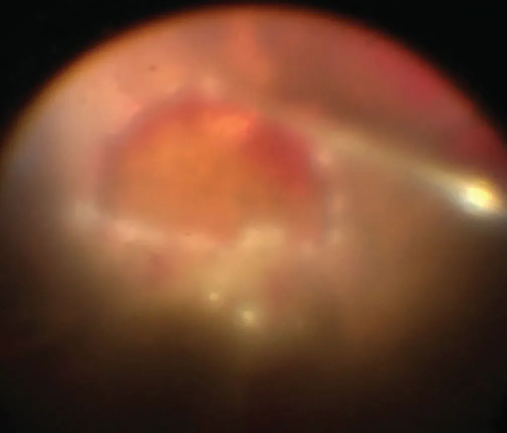
Once the break is addressed and traction is fully relieved, proceed with fluid-air exchange and laser retinopexy. Do not rush to close without confirming complete retinal apposition under the gas bubble.
MANAGING INTRAOPERATIVE BLEEDING
Bleeding is the most common complication and among the most anxiety-provoking. The approach is straightforward once internalized and includes the following steps:
- Raise the infusion pressure. Tamponade first, then immediately identify the source. Increasing IOP through the infusion line will slow most hemorrhages long enough to locate the culprit vessel.
- Apply diathermy. Always have bipolar diathermy primed and accessible at the start of every case. When a vessel bleeds, apply diathermy precisely and with minimal energy—just enough to achieve hemostasis.
- Perform a focal retinectomy. Rarely, a focal retinectomy may be necessary when there is a break and the subretinal hemorrhage becomes fused with an intraretinal/preretinal hemorrhage and a fibrosis/stiff retina, preventing release of traction from the break.
- Add perfluorocarbon liquid (PFCL). Although rarely needed, this can be useful in complex cases involving subretinal hemorrhage or a posterior break encountered while working anteriorly. Surgeons must remove the PFCL fully and ensure it remains in a single large bubble, so it does not enter subretinal space, given the posterior traction if there are any breaks.
END-OF-CASE DECISIONS
Once the membranes are cleared and the retina is flat, end-of-case decision making remains critical to durable anatomic success. Laser retinopexy should be applied to all breaks and in zones of peripheral ischemia. Additional panretinal photocoagulation delivered at the time of surgery is important in patients with limited follow-up compliance.
An intraoperative anti-VEGF injection should be strongly considered at case completion, particularly when residual neovascularization is visible or when the preoperative injection window was too short for full regression. This reduces the risk of postoperative hemorrhage and early reactivation.
Intravitreal or periocular steroids at case closure can suppress the postoperative inflammatory cascade, which can be robust in patients with poorly controlled diabetes.
Tamponade selection should favor gas over silicone oil in most complex cases. Long-acting gas (eg, C3F8 12% to 14%) provides 6 to 8 weeks of tamponade, avoids the need for oil removal surgery, and is associated with lower rates of secondary glaucoma and band keratopathy.4 Eyes receiving C3F8 may have a higher likelihood of visual improvement compared with those receiving silicone oil.5 Reserve silicone oil for cases involving a giant retinal tear, inferior retinectomy, poor patient positioning compliance, or when simultaneous bilateral surgery is planned.
PLAN CAREFULLY, BUT STAY FLEXIBLE
Complex diabetic vitrectomy requires a disciplined framework applied consistently from the preoperative evaluation through the final tamponade decision. By approaching each case with structured planning, a well-prepared instrument setup, and clear contingency protocols for bleeding and iatrogenic breaks, surgeons can navigate even the most advanced diabetic eyes with confidence and consistency.
1. Skondra D. Managing the challenging diabetic TRD. Presented at Vit-Buckle Society, Annual Meeting; April 10, 2026; Las Vegas.
2. Rizzo S, Genovesi-Ebert F, Di Bartolo E, Vento A, Miniaci S, Williams G. Injection of intravitreal bevacizumab (Avastin) as a preoperative adjunct before vitrectomy surgery in the treatment of severe proliferative diabetic retinopathy (PDR). Graefes Arch Clin Exp Ophthalmol. 2008;246(6):837-842. doi.org/10.1007/s00417-008-0774-y
3. Wakabayashi T, Oshima Y. Limited membranectomy for diabetic TRD. Retina Today. 2025;20(3):49-50.
4. Shettigar MP, Dave VP, Chou HD, et al. Vitreous substitutes and tamponades - A review of types, applications, and future directions. Indian J Ophthalmol. 2024;72(8):1102-1111. doi.org/10.4103/IJO.IJO_2417_23
5. Sokol JT, Schechet SA, Rosen DT, Ferenchak K, Dawood S, Skondra D. Outcomes of vitrectomy for diabetic tractional retinal detachment in Chicago’s county health system. Plos One. 2019;14(8):e0220726. doi.org/10.1371/journal.pone.0220726


