



An 11-year-old girl presented with a sudden decrease in vision in her left eye for a week. She reported no history of trauma or positive family history. Her medical history included oral ulcers.
On examination, her BCVA was 20/80 OD and hand motion at 20 cm OS. The anterior segment examination revealed the presence of 2+ cellular reaction in each eye.
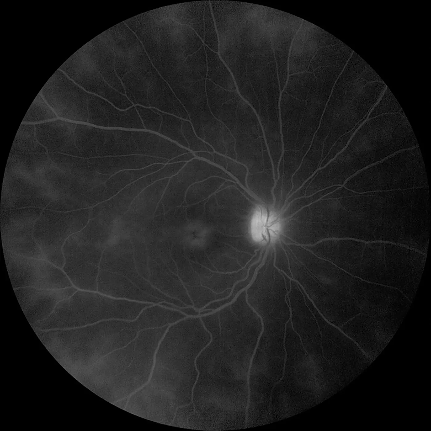
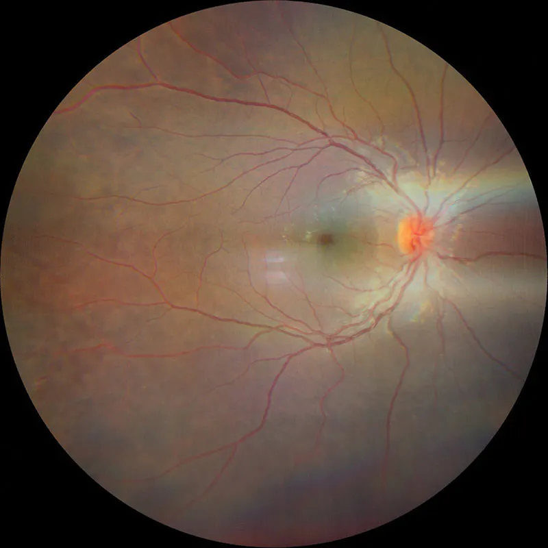
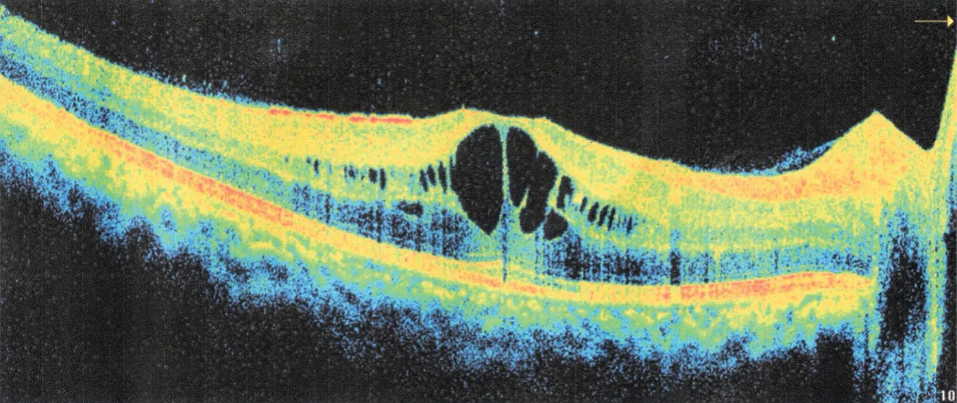
The fundus examination revealed the presence of vitreous hemorrhage in her left eye. Her right eye showed cystoid macular edema (CME) with active vasculitis. Fluorescein angiography in her right eye revealed the presence of hyper fluorescence of the disc, CME, and perivenular leakage in a fern-like pattern (Figure 1). Based on these findings and fundus and OCT imaging (Figures 2 and 3), she was diagnosed with Behçet disease.
The patient was started on systemic corticosteroids, received an intravitreal anti-VEGF injection in each eye, and was scheduled to receive panretinal photocoagulation in each eye before being lost to follow-up.
DISCUSSION
Behçet disease is an autoinflammatory disorder that predominantly affects the mucocutaneous, ocular, urogenital, vascular, and gastrointestinal systems. Ocular involvement occurs in nearly 70% of patients and often presents as recurrent bilateral nongranulomatous panuveitis and retinal vasculitis. It typically manifests between 20 and 40 years of age. Fluorescein angiography commonly reveals hyperfluorescence of the optic disc, vascular dye leakage from retinal capillaries, and staining of retinal vessels due to retinal perivasculitis. Diffuse vascular leakage in a fern-like pattern is the characteristic finding and is commonly observed during the chronic phases of inflammation.1-3
The main goal of treatment includes resolving the intraocular inflammation, preventing recurrent flare-ups, and achieving complete remission while preserving vision. Systemic corticosteroids, immunosuppressive drugs, and biologics are used.4 Panretinal photocoagulation along with anti-VEGF agents may be necessary to manage neovascularization secondary to chronic ischemic retinal vasculitis. The prompt and effective treatment of uveitis flare-ups, ensuring rapid resolution of intraocular inflammation, is essential to prevent vision loss.
1. Kaur M, Yip K. The current and novel imaging modalities for ocular vasculitis in Behçet’s disease: A review. Cureus. 2024;16(9):e69528.
2. Cunningham ET Jr, Tugal-Tutkun I, Khairallah M, Okada AA, Bodaghi B, Zierhut M. Behçet uveitis. Ocul Immunol Inflamm. 2017;25(1):2-6.
3. Zając H, Turno-Kręcicka A. Ocular manifestations of Behçet’s disease: An update on diagnostic challenges and disease management. J Clin Med. 2021;10(21):5174.
4. Khieu C, Chanthan D. Novel approach to Behçet’s disease in the era of biologic agents. Curr Opin Ophthalmol. 2023;34(6):535-542.
_1784132761.jpg?auto=compress,format&w=70)


